This campaign has ended.

How to get your order discounted 10% |
|
|
|
|
Direct orders to Dojindo and US customers onlySurvey expiration date: May. 24, 2024 |
|
How to get your order discounted 10% |
|
|
|
|
Direct orders to Dojindo and US customers onlySurvey expiration date: May. 24, 2024 |
|
Following topics will be informed through Dojindo newsletter. For the registration of newsletter, please click here.
|
Ferroptosis is a form of programmed cell death characterized by the accumulation of lipid peroxides to lethal levels and is distinct from other forms of cell death such as apoptosis, necroptosis, and autophagy. In the context of cancer, ferroptosis may act as a tumor suppressor mechanism, as cancer cells often have an increased susceptibility to ferroptosis due to their altered metabolism and increased levels of reactive oxygen species (ROS). Therapeutically, inducing ferroptosis in cancer cells has emerged as a promising strategy for cancer treatment, particularly for tumors that are resistant to traditional therapies such as chemotherapy and radiation. In addition, understanding the specific vulnerabilities of cancer cells to ferroptosis may aid in the design of targeted therapies that exploit these weaknesses, providing a potential avenue to overcome drug resistance and improve patient outcomes. |
||||||||
|
Dietary restriction of cysteine and methionine sensitizes gliomas to ferroptosis and induces alterations in energetic metabolism |
Lysosomal cystine governs ferroptosis sensitivity in cancer via cysteine stress response |
Mitochondria regulate intracellular coenzyme Q transport and ferroptotic resistance via STARD7 |
||||||
|
Point of Interest - A cysteine-depleted, methionine-restricted diet can improve the therapeutic response to RSL3 and prolong survival in a syngeneic orthotopic murine glioma model. - This CMD diet profoundly alters in vivo metabolome, proteome, and lipidome. |
Point of Interest - A shortage of cystine in lysosomes stimulates ATF4 expression through the AhR signaling pathway. - A weakened cysteine stress response increases sensitivity to ferroptosis during cysteine deprivation. - CysRx promotes cancer cell ferroptosis through intracellular nutrient reprogramming. |
Point of Interest - The mitochondrial STARD7 supports coenzyme Q synthesis, promotes oxidative phosphorylation, and maintains cristae morphology. - Its cytosolic counterpart facilitates the transport of coenzyme Q to the plasma membrane and protects against ferroptosis. - Overexpression of cytosolic STARD7 increases the resistance of cells to ferroptosis and reduces the availability of coenzyme Q in the mitochondria. |
||||||
| Related Techniques | ||||||||
| Intracellular / mitochondrial lipid peroxidation detection | Liperfluo, MitoPeDPP | |||||||
| Intracellular / mitochondrial ferrous ion (Fe2+) detection | FerroOrange, Mito-FerroGreen | |||||||
| Mitochondrial superoxide detection | MitoBright ROS Deep Red - Mitochondrial Superoxide Detection | |||||||
| Oxygen consumption rate assay | Extracellular OCR Plate Assay Kit | |||||||
| Lysosomal function | Lysosomal Acidic pH Detection Kit-Green/Red and Green/Deep Red NEW | |||||||
| Glycolysis/Oxidative phosphorylation Assay | Glycolysis/OXPHOS Assay Kit | |||||||
| Related Applications | ||||||||
|
||||||||
|
Autophagy is a cellular process that degrades and recycles damaged organelles, misfolded proteins, and other cellular debris, and is critical for maintaining cellular homeostasis. Dysregulated autophagy is often observed in the context of neurodegeneration, where either excessive or insufficient autophagic activity can contribute to the accumulation of toxic proteins and organelle dysfunction that are hallmarks of neurodegenerative diseases. Enhancing autophagy has been shown to alleviate symptoms and pathology in models of diseases such as Alzheimer's, Parkinson's, and Huntington's, suggesting a protective role against neurodegeneration. Conversely, excessive autophagy can lead to cell death, suggesting that a delicate balance is required for neuronal health and highlighting the complexity of targeting autophagy as a therapeutic strategy for neurodegenerative diseases. |
|||||
| Autophagy enables microglia to engage amyloid plaques and prevents microglial senescence Click here for the original article: Insup Choi, et. al., Nature Cell Biology, 2023. |
Microglial-to-neuronal CCR5 signaling regulates autophagy in neurodegeneration Click here for the original article: Beatrice Paola Festa, et. al., Neuron, 2023. |
Mitochondrial control of microglial phagocytosis by the translocator protein and hexokinase 2 in Alzheimer’s disease Click here for the original article: Lauren H. Fairley, et. al., PNAS, 2022. |
|||
|
Point of Interest - An autophagy deficiency fosters the development of senescent microglia. - Pharmacological removal of autophagy-deficient senescent microglia can alleviate neuropathology in mice with AD. |
Point of Interest - CCL-5, CCL-4, and CCL-3 derived from microglia activate the neuronal CCR5 receptor, leading to the inhibition of neuronal autophagy. - Levels of CCR5, CCL-3, CCL-4, and CCL-5 are elevated in the brains of mice with Huntington’s disease and tauopathy. - Inhibiting CCR5, either pharmacologically or genetically, ameliorates neurodegeneration in mice. |
Point of Interest - Hexokinase-2 (HK) affects glycolytic metabolism and phagocytosis through its interaction with mitochondria. - Mitochondrial HK influences glycolysis and inflammation, and its displacement improves phagocytosis in TSPO-deficient microglia. - Alzheimer’s beta-amyloid drastically stimulated mitochondrial HK recruitment in cultured microglia, which may contribute to microglial dysfunction in Alzheimer’s disease. |
|||
| Reported Examples: Autophagy and Mitophagy Detection Probes | |||||
| Detection (Reagent) | Samples | Reference | |||
| Autophagosome/Autolysosome (Autophagic Flux Assay Kit) |
Immortalized Fischer rat Schwann cells | International Journal of Molecular Sciences | |||
|
Autolysosome |
Cortical neurons | Neurobiology of Disease | |||
| PC12 cells(a mixture of neuroblastic cells and eosinophilic cells) | Hindawi | ||||
| Autophagosome (DAPGreen or DAPRed) |
Neurons from mouse embryonic stem cells | Life Science Alliance | |||
| Hippocampal neurons | PNAS | ||||
| N2a cells (Mouse Albino neuroblastoma) | International Journal of Molecular Sciences | ||||
| Mitophagy (Mtphagy Dye) |
Neural progenitor cell (NPC) | International Journal of Molecular Sciences | |||
| Primary cortical neuronal (CxN) cells | Cells | ||||
| Brain slices (mice) | The FEBS Journal | ||||
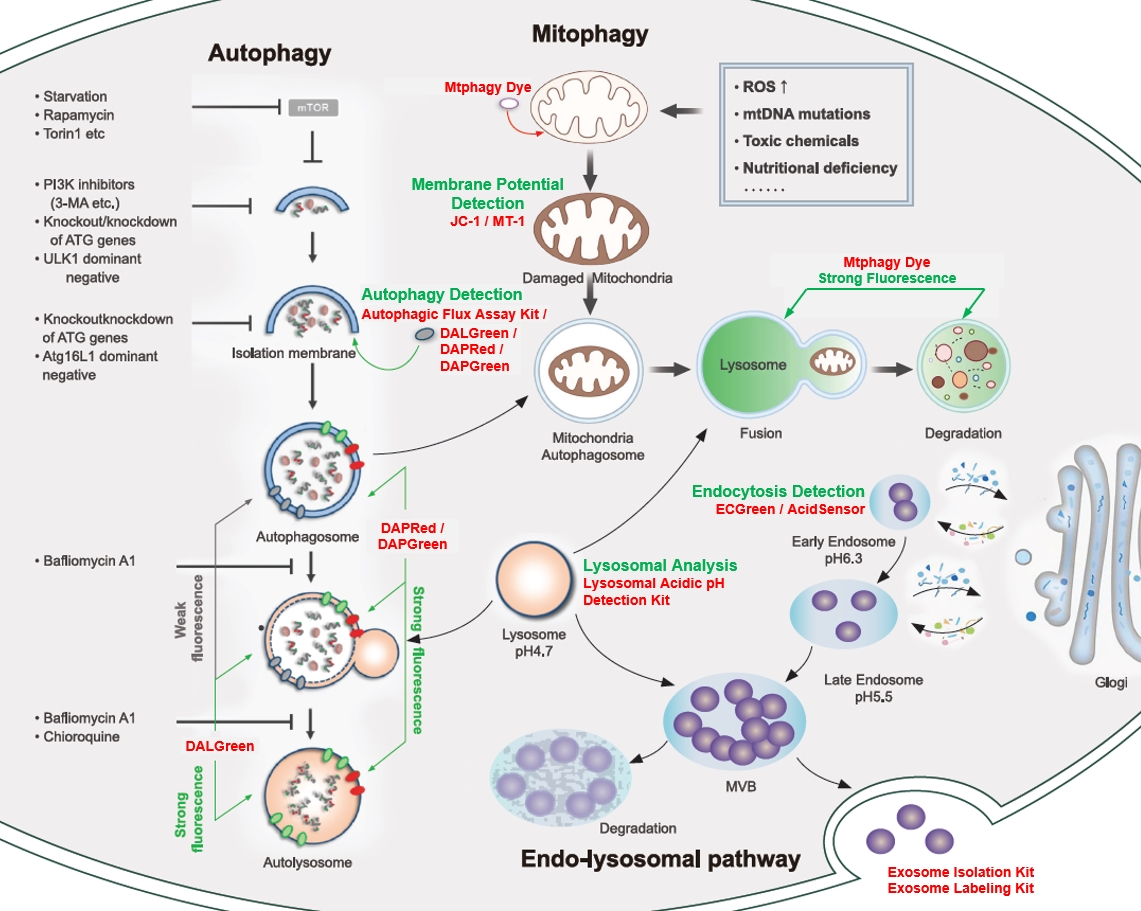
<Cellular Landscape: autophagy-related pathway>
|
Mitophagy Detection Mitochondrial Membrane Potential Detection Lysosomal Analysis Endocytosis Detection Exosome |
||||
|
Senescence is a cellular process that results in the cessation of cell division, often serving as a protective mechanism against the proliferation of damaged cells, including potential cancer cells. This process is intricately regulated by numerous factors including, but not limited to, tumor suppressor genes, DNA damage response (DDR) pathways, and various signaling molecules. In addition, the senescence-associated secretory phenotype (SASP), consisting of cytokines, growth factors, and proteases, is regulated by NF-κB and other transcription factors that influence the tissue microenvironment and impact aging and disease processes. |
|||
|
HKDC1, a target of TFEB, is essential to maintain both mitochondrial and lysosomal homeostasis, preventing cellular senescence |
Iron accumulation drives fibrosis, senescence and the senescence-associated secretory phenotype Click here for the original article: Mate Maus, et. al., Nature, 2023. |
Microautophagy regulated by STK38 and GABARAPs is essential to repair lysosomes and prevent aging Click here for the original article: Monami Ogura, et. al., EMBP Reports, 2023. |
|
|
Point of Interest - This activity helps avert cellular senescence, playing a vital role in maintaining cellular homeostasis. - Beyond its role in glycolysis, HKDC1 contributes to mitophagy and lysosomal repair processes independently. - The absence of HKDC1 may result in cellular senescence and the buildup of damaged organelles.
|
Point of Interest - Senescent cells persistently accumulate iron, even after the increase in extracellular iron has subsided. - Cells exposed to various types of senescence-inducing insults accumulate abundant ferritin-bound iron, mostly within lysosomes. |
Point of Interest - Microautophagy in the repair of damaged lysosomes prevents aging. - STK38 and GABARAPs are key regulators of this process. - STK38 is required for lysosomal recruitment of VPS4 and GABARAPs are involved in ESCRT assembly. - Depletion of these regulators leads to accelerated cellular senescence and shortened lifespan. |
|
| Related Techniques | |||
| Cellular senescence detection | SPiDER-βGal for live-cell imaging or flow cytometry / microplate reader / tissue samples. | ||
| First-time autophagy research | Autophagic Flux Assay Kit | ||
| Autophagy detection | DAPGreen / DAPRed (Autophagosome detection), DALGreen (Autolysosome detection) | ||
| Lysosomal function | Lysosomal Acidic pH Detection Kit-Green/Red and Green/Deep Red NEW | ||
| Ferrous ion (Fe2+) detection | FerroOrange(intracellular), Mito-FerroGreen(mitochondria) | ||
| Mitochondrial superoxide detection | MitoBright ROS Deep Red - Mitochondrial Superoxide Detection | ||
| Oxygen consumption rate assay | Extracellular OCR Plate Assay Kit | ||
| Related Applications | |||
Analysis of Lysosomal Mass and pH Exchange in Senescence-induced Cells |
|||
|
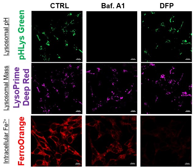
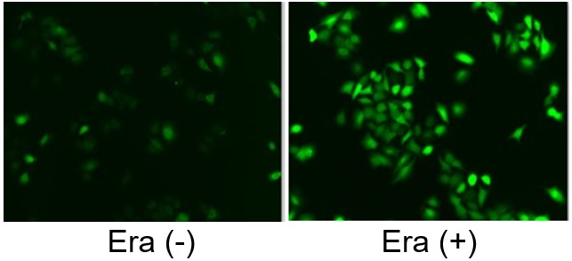
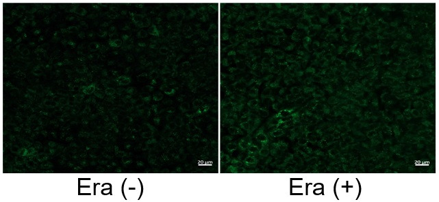
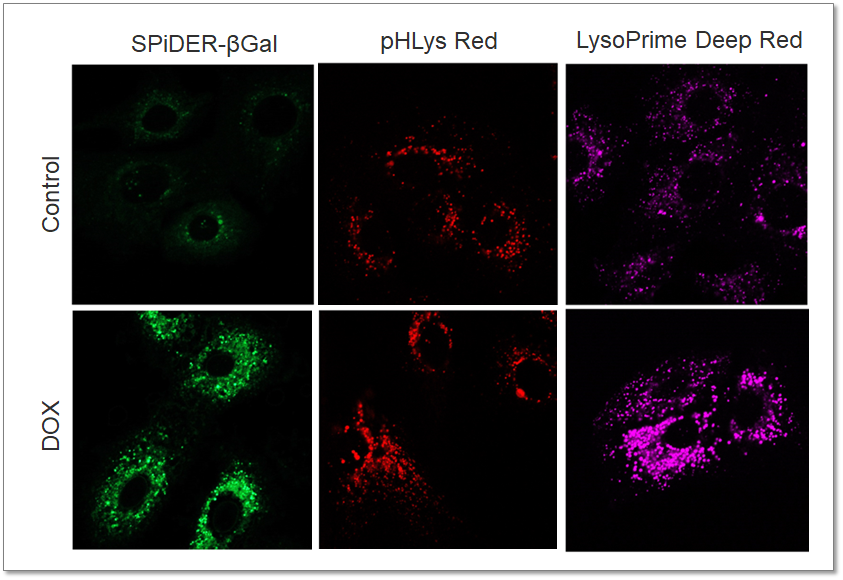
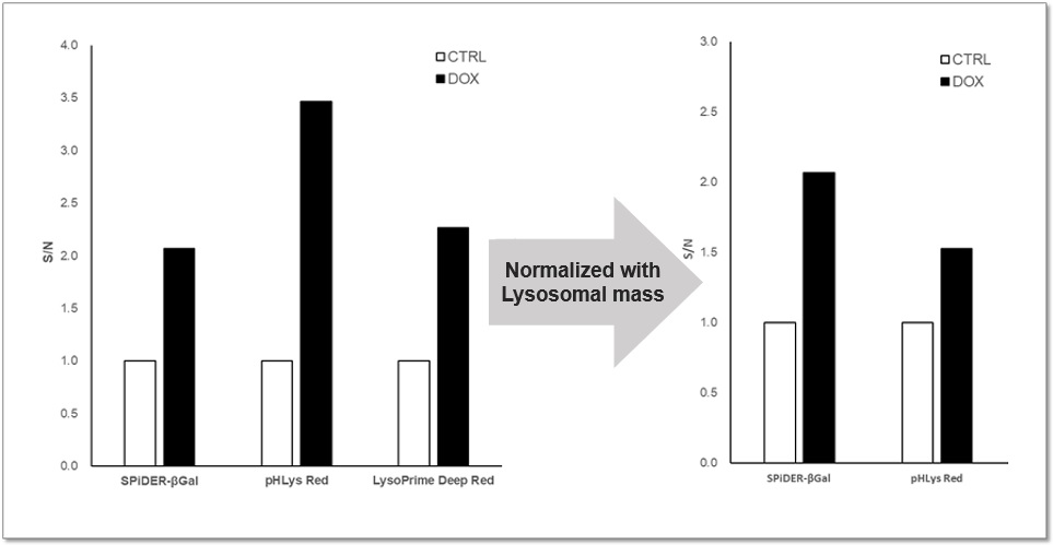
Purpose: To investigate changes in lysosomal mass and pH in A549 cells induced to senescence by treatment with Doxorubicin (DOX). Methods: Senescence-associated acidic β-galactosidase (SA-βGal) activity was detected using Cellular Senescence Detection Kit - SPiDER-βGal. Lysosomal mass was detected using LysoPrime Deep Red, and pH was detected using pHLys Red. Fluorescence imaging was used to observe changes in lysosomal mass and pH in senescent cells compared to non-senescent cells. The normalized fluorescence intensity of lysosomal mass and pH was also measured by a plate reader. Results: Our findings indicate that senescence induced by DOX resulted in an increase in lysosomal mass and acidification of pH compared to non-senescent cells. The obtained results are consistent with previous reports* that demonstrated enhanced lysosomal activity in senescent cells induced by the CDK4/6 inhibitor, palbociclib. The fluorescence imaging and plate reader data both support these findings. * Miguel Rovira, et. al., Aging Cell (2022) <Experimental Conditions for Microscopy> <Experimental Conditions for Plate Reader> <Products in Use> |
|
||
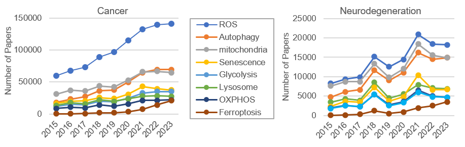
Trends in various indices of cell function analysis in terms of number of papers (referenced from Dimensions)
Dojindo categories and products are ranked based on sales, online searches, and inquiries over the past six months.
| Ranking | Category | Product (Catalog Page#) |
|---|---|---|
| 1 |
Ferroptosis |
FerroOrange (14), Liperfluo (12), Cystine Uptake Assay Kit (21) |
| 2 |
Autophagy |
|
| 3 |
Glycolysis/OXPHOS |
|
| 4 | Senescence | Cellular Senescence Detection Kit – SPiDER-ßGal (6) |
| 5 |
Mitochondria |
Mitophagy Detection Kit (26), MitoBright ROS Deep Red (27) |
| 6 |
Lysosome |
|
| 7 |
ROS |
|
| 8 |
Lipid |
| Category | Product Name | Feature |
|---|---|---|
| Cell Proliferation / Cytotoxicity Assay | Cell Counting Kit-8 Best Seller | Simple-process cell viability assay using metabolism (dehydrogenase activity) as an indicator |
| Cytotoxicity LDH Assay Kit-WST | Reliable cytotoxicity assay by released LDH activity | |
| Cell Count Normalization Kit | Normalization of cell counts by nuclear staining | |
| Viability/Cytotoxicity Multiplex Assay Kit NEW | Set of Cell Counting Kit-8 and Cytotoxicity LDH Assay Kit-WST (500 tests each) | |
| Senescence | Cellular Senescence Detection Kit – SPiDER-ßGal HOT | First choice for cellular senescence assay |
| SPiDER-ßGal | SA-β-gal assay for living cells and tissue | |
| Cellular Senescence Plate Assay Kit – SPiDER-ßGal | Cellular senescence assay with a plate reader | |
| Nucleolus Bright Green | Nucleolus fluorescent staining | |
| Nucleolus Bright Red | ||
| Ferroptosis | Liperfluo HOT | Intracellular lipid peroxide measurement |
| MitoPeDPP | Mitochondria lipid peroxidation measurement | |
| Mito-FerroGreen | Mitochondria ferrous ion (Fe2+) detection | |
| FerroOrange HOT | Intracellular ferrous ion (Fe2+) detection | |
| Lipid Peroxidation Probe -BDP 581/591 C11- | Lipid peroxidation detection | |
| ROS / Oxidative Stress | GSSG/GSH Quantification Kit | Quantification of reduced and oxidized glutathione |
| ROS Assay Kit - Highly Sensitive DCFH-DA - NEW | Higher sensitivity than conventional DCFH-DA | |
| ROS Assay Kit - Photo-oxidation Resistant DCFH- DA - NEW | Reduced photo-oxidation and compatible with fixation | |
| MitoBright ROS Deep Red - Mitochondrial Superoxide Detection NEW | Mitochondrial superoxide detection by deep red staining, co-staining with other markers | |
| Lysosome | Lysosomal Acidic pH Detection Kit-Green/Red NEW | Ready-to-use kits for Lysosomal pH and mass detection |
| Lysosomal Acidic pH Detection Kit-Green/Deep Red NEW | ||
| pHLys Red - Lysosomal Acidic pH Detection NEW | Highly sensitive lysosomal pH detection dye | |
| LysoPrime Green / Deep Red – High Specificity and pH Resistance NEW | Accurate lysosome detection with high selectivity and resistance to pH changes | |
| Cellular Metabolism | ATP Assay Kit-Luminescence | Easy quantification of intracellular ATP with a plate reader |
| Glucose Assay Kit-WST (Colorimetric) | Easy quantification of various intracellular or extracellular indicators with a plate reader | |
| Glutamine Assay Kit-WST (Colorimetric) | ||
| Glutamate Assay Kit-WST (Colorimetric) | ||
| Glycolysis/OXPHOS Assay Kit NEW | ||
| Glycolysis/JC-1 MitoMP Assay Kit NEW | ||
| Lactate Assay Kit-WST (Colorimetric) | ||
| α-Ketoglutarate Assay Kit-Fluorometric | Easy quantification of various intracellular indicators with a plate reader | |
| NAD/NADH Assay Kit-WST (Colorimetric) | ||
| NADP/NADPH Assay Kit-WST (Colorimetric) | ||
| Extracellular OCR Plate Assay Kit HOT NEW | Oxygen Consumption Rate (OCR) detection with a fluorescent plate reader | |
| Nutrient Uptake | Glucose Uptake Assay Kit- Blue / Green / Red | Highly sensitive and easy to measure each indicator with a fluorescent plate reader without RI-labeled |
| Amino Acid Uptake Assay Kit NEW | ||
| Cystine Uptake Assay Kit NEW | ||
| Fatty Acid Uptake Assay Kit NEW | ||
| Mitochondrial Staining | MitoBright LT Green / Red / Deep Red | Mitochondria imaging with long-term visualization |
| MitoBright IM Red for Immunostaining | Mitochondria imaging with capable of co-staining with immunomarkers | |
| mtSOX Deep Red – Mitochondrial Superoxide Detection NEW | Mitochondrial superoxide detection by deep red staining, co-staining with other markers | |
| MitoPeDPP | Mitochondria lipid peroxidation measurement | |
| Mito-FerroGreen | Mitochondria ferrous ion (Fe2+) detection | |
| Mitochondrial Membrane Potential | JC-1 MitoMP Detection Kit | Widely used and many papers reported |
| MT-1 MitoMP Detection Kit NEW | More sensitive than JC-1 and fixable after staining | |
| Mitophagy | Mitophagy Detection Kit | First choice for mitophagy detection |
| Mtphagy Dye | For experienced users of the mitophagy detection kit | |
| Lipid Droplet | Lipi-Blue / Green / Red / Deep Red | Accurate lipid droplet imaging using living and PFA fixed cells with low background |
| Lipid Droplet Assay Kit-Blue / Deep Red | Accurate lipid droplet measurement with plate reader and flow cytometer | |
| Autophagy | DALGreen - Autophagy Detection HOT | Selective autolysosome detection in live cells |
| DAPGreen - Autophagy Detection HOT | Selective autophagosome detection in live cells | |
| DAPRed - Autophagy Detection HOT | ||
| Autophagy Flux Assay Kit NEW | First-choice product for autophagy research | |
| Endocytosis | ECGreen-Endocytosis Detection NEW | Accurate detection of endocytosis by pH changes |
| AcidSensor Labeling Kit – Endocytic Internalization Assay NEW | Labeling pH-sensitive fluorescent dye to antibody or protein to monitor the endocytosis uptake | |
| Phagocytosis | AcidSensor Labeling Kit – Endocytic Internalization Assay | Detect phagocytosis activity with flow cytometry. The application is available on the product page. |
| Exosome | ExoIsolator Exosome Isolation Kit NEW | Exosomes are obtained quickly without any complicated operations from a supernatant |
| ExoIsolator Isolation Filter NEW | ||
| ExoSparkler Exosome Membrane Labeling Kit-Green / Red / Deep Red | Exosome membrane – fluorescent labeling (highly precise) | |
| Plasma Membrane | PlasMem Bright Green / Red | Cell membrane staining reagents with low cytotoxicity |
Cell Proliferation / Cell Cytotoxicity Assay Kits /Related Reagents
Cell Double Staning Kit /Live Cell Staining /Dead Cell Staining /Nuclear Staining /Mitochondria Staning /Tissue Staining /Nucleolus Staining /Lipid Droplet Staining /Cell Membrane Staining /Lysosome Staining
Reagents for Intracellular Calucuum Ion /Reagents for Intracellular Ion /Related Reagents
Protein Labeling Kits /Protein Labeling Reagents /HPLC Derivertization Reagents /Biotion Labeling Reagents /Related Reagents /Exsosome Labeling
Stress Maker Detection /NO Detection /NO Donor /NO Inhibitor /ACE Inhibition Assay /Reagents・Kits for Sulfur Biology /Antioxidant Assay Kit /Donors for Sulfur Biology
Bacterial Proliferation Assay Kit /Bacteria Staining /Bacterial Fluorescent Staining
Transfection Reagents /Nuclear Staining /Agarose /Related Reagents /Buffer for Molecular Biology
Detergents /Sets
Hetero-bifunctional Reagents /Homo-bifunctional Reagents /Others
Reductive Chromogenic Dyes /Electron Mediators /Oxidative Chromogenic Dyes /Trinder Reagents
Ionophores /Anion Eliminator /Solvent for Ion Electrode of Liquid Film Type
Buffers
EDTA /Other Chelator /Reagents for Chelator Titration
Chromogen/Metal Indicator
/Fluorine /Iron / /Water Hardness /Residual Chlorine /ABS /Cyan / /Chromium /Copper
AA Chelator /Related Reagents
Spectrozole /Luminazole / /Acnazole /Dehydration Solvent for Synthesis
Biochemicals
Alkanethiol Derivative /Phosphonic Acid Derivatives