Lipotoxicity is caused by intracellular lipid accumulation and is indicative of mitochondrial disfunction. Lipotoxicity accelerates the degenerative process of cellular senescence, influencing cancer development.

References
- 1. Clara, C. al., “Mitochondria: Are they causal players in cellular senescence?”, Biochimica et Biophysica Acta – Bioenergetics, 2015, 1847(11), 1373-1379.
- 2. Huizhen, Z. et al., “Lipidomics reveals carnitine palmitoyltransferase 1C protects cancer cells from lipotoxicity and senescence”, Journal of Pharmaceutical Analysis, 2020.
- 3. Xiaojuan, H. et al., “Astrocyte Senescence and Alzheimer’s Disease: A Review”, Front. Aging Neurosci., 2020.
- 4. Borén, J. et al., “Apoptosis-induced mitochondrial dysfunction causes cytoplasmic lipid droplet formation”, Cell Death Differ, 2012, 19(9), 1561-1570.
- 5. Na, L. et al., “Aging and stress induced β cell senescence and its implication in diabetes development”, Aging (Albany NY), 2019, 11(21), 9947–9959.
① Lipid Droplet Detection
The Lipi-series kits can clearly identify lipid droplets with an extremely low level of background fluorescence and specificity better than that of BODIPY 493/503 or Nile Red.
We observed lipid droplet generation in young and aged mice using mouse liver adipose tissue (frozen sections).

Lipi-series kits are available in four colors (blue, green, red, and deep-red) and can be used for multiple stain analysis as shown in the following papers.
| No. |
Sample |
Multiple Staining |
Reference |
| 1 |
Brown Adipocytes |
Co-stained with MitoBright Green (Mitochondria), Hoechst 33342 (Nuclear) and Lipi-Red |
Masako, O. et al., “Exogenous Cytokine-Free Differentiation of Human Pluripotent Stem Cells into Classical Brown Adipocytes”, Cells., 2019, 8(4), 373. |
| 2 |
hMGEC |
Co-stained with tandem RFP-GFP-tagged LC3B (autophagosome /autophagolysosome) and Lipi-Blue |
Kim, S. et al., “Eicosapentaenoic acid (EPA) activates PPARγ signaling leading to cell cycle exit, lipid accumulation, and autophagy in human meibomian gland epithelial cells (hMGEC)”, The Ocular Surface, 2020, 18(3), 427-437. |
| 3 |
AdMSCs |
Co-stained with UCP1 antibody (Alexa Fluor 488), DAPI (Nuclear) and Lipi-Red |
Yukimasa, T. et al., “Transcriptome analysis reveals brown adipogenic reprogramming in chemical compound-induced brown adipocytes converted from human dermal fibroblasts”, Scientific Reports, 2021, 11, 5061. |
Related Products
② Mitochondria Dynamics
Abnormalities in the morphology and dynamics of mitochondria have been associated with cellular senescence. This association has inspired a demand to be able to observe mitochondrial dynamics over a long period.
Fortunately, MitoBright LT has been confirmed as being retained in mitochondria even after 4 days in HeLa cells.

Related Products
③ Senescence Marker Detection
Cellular Senescence Detection Kit – SPiDER-βGal allows for detection of Senescence-Associated-β-Galactosidase (SA-β-gal) with high sensitivity and ease-of-use. SPiDER-βGal is a new reagent for detecting β-galactosidase, possessing high cell permeability and high retentivity inside cells. SA-β-gal can be detected specifically in either fixed or living cells by using Bafilomycin A1 to inhibit endogenous β-galactosidase activity.
We used SPiDER-βGal to analyze the associations between cellular senescence, mitochondrial membrane potential, and cell cycle phase proportions. Doxorubicin (DOX) acts to inhibit cell proliferation during G2/M phases of the cell cycle and induces cellular senescence. After adding DOX to A549 cells, higher histogram peaks for the G2/M phase (Cell Cycle Assay Solution Blue and Deep Red), elevated senescence markers (Cellular Senescence Detection Kit – SPiDER-βGal), and changes in mitochondrial membrane potential (JC-1 MitoMP Detection Kit) were observed.

④ Monitoring Mitochondrial Membrane Potential
JC-1, TMRE, and TMRM are widely used to monitor mitochondrial membrane potential. However, these dyes have limitations, such as low photostability and poor retention after aldehyde fixation. These limitation result in experiments with poor reproducibility.
MT-1 MitoMP Detection Kit has high photostability and remains unquenched even in cells that have been fixed with paraformaldehyde after staining. These features allow the MT-1 Kit to produce highly reproducible results.
In addition, the Imaging Buffer included in this kit minimizes background fluorescence and maintains cell vitality while the assay is being performed.

Related Products
⑤ Mitophagy Detection
The Mitophagy Detection Kit does not require protein expression/transfection. Mitophagy can be easily detected by simply adding reagents.

The association between cellular senescence and mitophagy can be confirmed using the Mitophagy Detection Kit.
Arsun, B. at al., “Age-associated changes in human CD4+ T cells point to mitochondrial dysfunction consequent to impaired autophagy“, Aging (Albany NY), 2019, 11(21), 9234–9263.
As well, in order to investigate the influence and involvement of mitochondria in lipofuscinogenesis, Dr. König et al. analyzed lipofuscin amounts as well as mitophagy in senescent cells.
Jeannette, K. at al., “Mitochondrial contribution to lipofuscin formation“, Redox Biol., 2017, 11, 673–681.
Related Products
Lysosome Detection
Many types of small fluorescent probes are used for monitoring lysosomes in living cells. Dojindo’s LysoPrime Green overcomes known problems with fluorescent lysosome probes, such as lack of specificity for lysosomes and staining dependent on the lysosomal pH. In addition, the high-retentivity of LysoPrime Green enables long-term imaging experiments.

LysoPrime Green staining had retained inside the lysosome even during the long periods of stimulation required for autophagy and mitophagy induction.
Related Products
⑥ Lipid Peroxide Detection
Liperfluo is a fluorescent probe that can directly detect intracellular lipid peroxide. Lipid peroxidation plays a critical role in the specific form of cell death known as ferroptosis. A great amount of research in recent years has focused on ferroptosis.
Brent, S. et al., “Ferroptosis: A Regulated Cell Death Nexus Linking Metabolism, Redox Biology, and Disease“, Cell, 2017, 171(2), 273.
Valerian, K. al., “Oxidized Arachidonic/Adrenic Phosphatidylethanolamines Navigate Cells to Ferroptosis“, Nat. Chem. Biol., 2017, 13(1), 81.

*Little is currently known about the morphology and/or distribution of lipid droplets during ferroptosis
Related Products
⑦ Glucose Uptake Detection
Glucose Uptake Assay Kit-Green allows for highly sensitive detection of cellular glucose uptake via fluorescence microscopy, flow cytometry, or microplate assay.
Unlike fluorescent dye 2-NBDG, which decreases in fluorescence intensity in water, this kit uses a fluorescent dye with superior brightness.

Related Products
⑧ Total ROS Detection
DCFH-DA is widely used for ROS detection, but it has some limitations like weak fluorescence signals and high background. ROS Assay Kit -Highly Sensitive DCFH-DA- was developed to correct these limitations. The dye employed in the kit allows ROS detection with higher sensitivity than DCFH-DA.

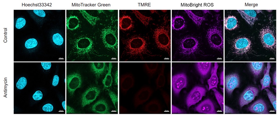
Mitochondrial Superoxide Detection
MitoSOX™ Red has widely been used to detect mitochondrial superoxide. However, the emission wavelength is the common red, which overlaps with other MMP detection probes such as TMRE, and is therefore not applicable for simultaneous staining with these other mitochondrial markers in a single sample.
Dojindo’s MitoBright ROS Deep Red overcomes this limitations. This dye emits deep red fluorescence; its fluorescence does not overlap with emission wavelengths that other red fluorescent markers use. Furthermore, the MitoBright ROS Deep Red is better able to selectively detect superoxide, compared to MitoSOX ™ Red.
⑨ Cell Cycle Analysis
Propidium iodide (PI) is generally used for cell cycle analysis via flow cytometry. As opposed to the PI method, Cell Cycle Assay Solution Blue/Deep Red has better membrane permeability and uses a dye with high specificity for DNA. These differences make it possible to analyze the cell cycle just by adding the reagent to a cell suspension.
Additionally, Cell Cycle Assay Solution Blue/Deep Red allows you to concurrently use the highly versatile 488 nm laser wavelength.

Related Products


