


Science Note: Ferroptosis
Cancer Ferroptosis: Relationship on Metabolism, Lysosome, and Mitochondria [Mar. 12, 2024]
Open / Close the Article
|
Ferroptosis is a form of programmed cell death characterized by the accumulation of lipid peroxides to lethal levels and is distinct from other forms of cell death such as apoptosis, necroptosis, and autophagy. In the context of cancer, ferroptosis may act as a tumor suppressor mechanism, as cancer cells often have an increased susceptibility to ferroptosis due to their altered metabolism and increased levels of reactive oxygen species (ROS). Therapeutically, inducing ferroptosis in cancer cells has emerged as a promising strategy for cancer treatment, particularly for tumors that are resistant to traditional therapies such as chemotherapy and radiation. In addition, understanding the specific vulnerabilities of cancer cells to ferroptosis may aid in the design of targeted therapies that exploit these weaknesses, providing a potential avenue to overcome drug resistance and improve patient outcomes. |
||||||||
|
Dietary restriction of cysteine and methionine sensitizes gliomas to ferroptosis and induces alterations in energetic metabolism |
Lysosomal cystine governs ferroptosis sensitivity in cancer via cysteine stress response |
Mitochondria regulate intracellular coenzyme Q transport and ferroptotic resistance via STARD7 |
||||||
|
Point of Interest - A cysteine-depleted, methionine-restricted diet can improve the therapeutic response to RSL3 and prolong survival in a syngeneic orthotopic murine glioma model. - This CMD diet profoundly alters in vivo metabolome, proteome, and lipidome. |
Point of Interest - A shortage of cystine in lysosomes stimulates ATF4 expression through the AhR signaling pathway. - A weakened cysteine stress response increases sensitivity to ferroptosis during cysteine deprivation. - CysRx promotes cancer cell ferroptosis through intracellular nutrient reprogramming. |
Point of Interest - The mitochondrial STARD7 supports coenzyme Q synthesis, promotes oxidative phosphorylation, and maintains cristae morphology. - Its cytosolic counterpart facilitates the transport of coenzyme Q to the plasma membrane and protects against ferroptosis. - Overexpression of cytosolic STARD7 increases the resistance of cells to ferroptosis and reduces the availability of coenzyme Q in the mitochondria. |
||||||
| Related Techniques | ||||||||
| Intracellular / mitochondrial lipid peroxidation detection | Liperfluo, MitoPeDPP | |||||||
| Intracellular / mitochondrial ferrous ion (Fe2+) detection | FerroOrange, Mito-FerroGreen | |||||||
| Mitochondrial superoxide detection | MitoBright ROS Deep Red - Mitochondrial Superoxide Detection | |||||||
| Oxygen consumption rate assay | Extracellular OCR Plate Assay Kit | |||||||
| Lysosomal function | Lysosomal Acidic pH Detection Kit-Green/Red and Green/Deep Red | |||||||
| Glycolysis/Oxidative phosphorylation Assay | Glycolysis/OXPHOS Assay Kit | |||||||
| Related Applications | ||||||||
|
||||||||


Ferroptosis Activation, Inhibition, and Sensitization [Feb. 13, 2024]
Open / Close the Article
|
Ferroptosis is a form of programmed cell death characterized by the accumulation of lipid peroxides to lethal levels and is distinct from other forms of cell death such as apoptosis or necrosis. In cancer, ferroptosis can act as a double-edged sword: on the one hand, inducing ferroptosis in cancer cells may be a promising therapeutic strategy, as many cancers are resistant to traditional forms of cell death such as apoptosis. On the other hand, cancer cells can develop resistance to ferroptosis, which contributes to therapy resistance and tumor progression. Understanding and manipulating the pathways that regulate ferroptosis in cancer cells holds the potential for developing novel cancer treatments and overcoming resistance to existing therapies. |
||||||||
|
Targeted activation of ferroptosis in colorectal cancer via LGR4 targeting overcomes acquired drug resistance |
7-Dehydrocholesterol is an endogenous suppressor of ferroptosis |
Sensitization of cancer cells to ferroptosis coincident with cell cycle arrest |
||||||
|
Point of Interest - Inhibition of LGR4-Wnt signaling sensitized drug-induced ferroptosis. - LGR4-dependent Wnt signaling upregulated SLC7A11, a key inhibitor of ferroptosis, leading to drug resistance. |
Point of Interest - On the other hand, 7-DHC accumulation confers a prosurvival function in cancer cells. - 7-DHC effectively protects (phospho)lipids from autoxidation and subsequent fragmentation due to its superior reactivity with peroxyl radicals. - Accumulation of 7-DHC induces a shift to a ferroptosis-resistant state in tumors. |
Point of Interest - Stabilization of p53 and inhibition of CDK4/6 decrease the expression of MBOAT1 and EMP2. - Loss of EMP2 increases cell sensitivity to GPX4 inhibitors by altering lipid metabolism. - An orally bioavailable GPX4 inhibitor shows in vivo activity. |
||||||
| Related Techniques | ||||||||
| Intracellular lipid peroxidation measurement | Liperfluo | |||||||
| Mitochondria lipid peroxidation measurement | MitoPeDPP | |||||||
| Intracellular ferrous ion (Fe2+) detection | FerroOrange | |||||||
| Mitochondria ferrous ion (Fe2+) detection | Mito-FerroGreen | |||||||
| Mitochondrial superoxide detection | MitoBright ROS Deep Red - Mitochondrial Superoxide Detection | |||||||
| Total ROS detection | Highly sensitive DCFH-DA or Photo-oxidation Resistant DCFH-DA | |||||||
| Lipid droplets detection | Lipi-Blue / Green / Red / Deep Red | |||||||
| Antibody/Protein labeling with fast and high recovery |
Fluorescein, Biotin, and Peroxidase Labeling Kit - NH2 | |||||||
| Related Applications | ||||||||
|
||||||||
Ferroptosis Induced by FSP1-dependent Phase Separation [July 25, 2023]
Open / Close the Article
|
Ferroptosis suppressor protein-1 (FSP1) has recently been identified as a second system that suppresses ferroptosis, preventing lipid peroxidation independently of the glutathione-GPX4 axis. Researchers have used a small molecule library screen to discover 3-phenylquinazolinones, represented by icFSP1, as potent inhibitors of FSP1. Unlike previous inhibitors, icFSP1 does not directly inhibit FSP1 enzyme activity, but causes FSP1 to relocate from the membrane and condense, working in synergy with GPX4 inhibition. Learn more about how the authors detected Lipid peroxide, one of the ferroptosis markers, using Liperfluo. We also offer ferroptosis-related products: FerroOrange, Mito-FerroGreen. |
|||||||||||||||||||
|
Phase separation of FSP1 promotes ferroptosis Click here for the original article: T. Nakamura, et. al., Nature (2023) Point of Interest |
|||||||||||||||||||
| Related Techniques | |||||||||||||||||||
| Intracellular lipid peroxidation measurement | Liperfluo | ||||||||||||||||||
| Mitochondria lipid peroxidation measurement | MitoPeDPP | ||||||||||||||||||
| Intracellular ferrous ion (Fe2+) detection | FerroOrange | ||||||||||||||||||
| Mitochondria ferrous ion (Fe2+) detection | Mito-FerroGreen | ||||||||||||||||||
| Mitochondrial superoxide detection | MitoBright ROS Deep Red - Mitochondrial Superoxide Detection | ||||||||||||||||||
| Total ROS detection | Highly sensitive DCFH-DA or Photo-oxidation Resistant DCFH-DA | ||||||||||||||||||
| Lipid droplets detection | Lipi-Blue / Green / Red / Deep Red | ||||||||||||||||||
| NAD(H) and NADP(H) redox couples assay | NAD/NADH and NADP/NADPH Assay Kit | ||||||||||||||||||
| Related Applications | |||||||||||||||||||
Erastin-Induced Ferroptosis: Evaluating Intracellular Uptake and Redox Balance
|
|||||||||||||||||||

Age-related Microglial Phenotype Characterized by Lipid and Iron Contents [July 5, 2023]
Open / Close the Article
|
Scientists have unveiled that in comparison to young mice, one-third of old microglia show Lipofustin-related autofluorescence (AF), characterized by profound changes in lipid and iron content, phagocytic activity, and oxidative stress. Pharmacological removal of microglia in older mice successfully eliminated AF-microglia, following the repopulation of new functional microglia leads to an improvement in age-related neurological impairments and reduces neurodegeneration after traumatic brain injury. Learn more about how the authors phenotyped AF-microglia using Lipi-Blue* for Lipid droplet labeling, and FerroOrange* for iron labeling. (Please refer to Fig. 1E, 5F, 9E for FerrOrange, Fig. 5D, 7E, for Lipi-Blue) *Listed at 2023 Summer Promotion! Click here. |
|||
|
Brain injury accelerates the onset of a reversible age-related microglial phenotype associated with inflammatory neurodegeneration Click here for the original article: Rodney M Ritzel, et. al., Sci Adv (2023) Point of Interest |
|||
| Related Techniques | |||
| Lipid droplets detection | Lipi-Blue / Green / Red / Deep Red | ||
| Intracellular ferrous ion (Fe2+) detection | FerroOrange | ||
| Mitochondria ferrous ion (Fe2+) detection | Mito-FerroGreen | ||
| Lysosomal function assay | Lysosomal Acidic pH Detection Kit-Green/Deep Red NEW | ||
| Lysosomal Acidic pH Detection Kit-Green/Red | |||
| Cellular senescence detection (Live cell imaging or FCM) | Cellular Senescence Detection Kit | ||
| Cellular senescence detection (Plate reader) | Cellular Senescence Plate Assay Kit | ||
| Mitochondrial superoxide detection | MitoBright ROS Deep Red - Mitochondrial Superoxide Detection | ||
| Total ROS detection | Highly sensitive DCFH-DA or Photo-oxidation Resistant DCFH-DA | ||
| Related Applications | |||
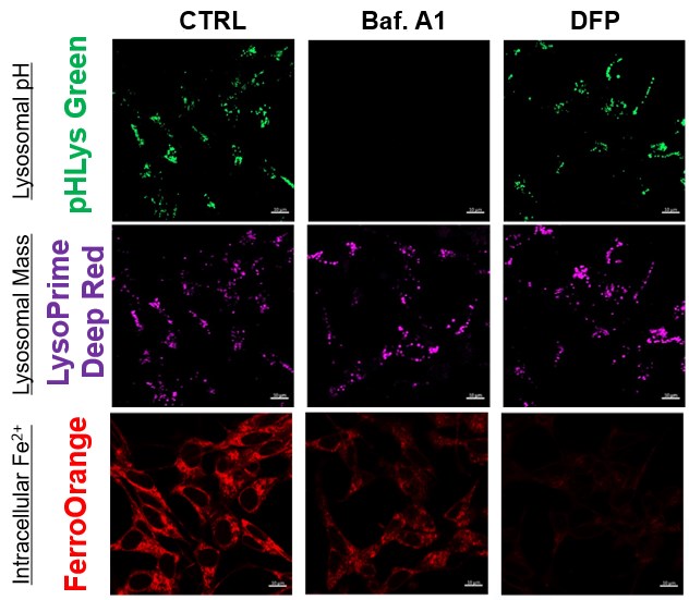
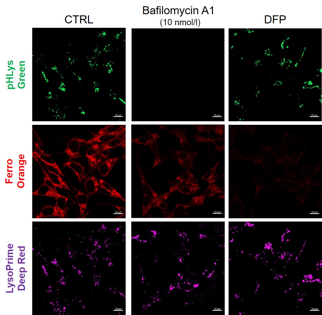
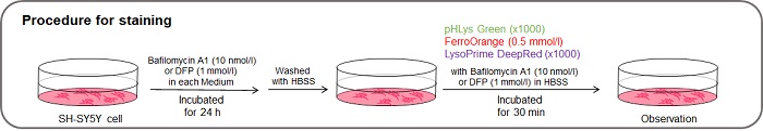
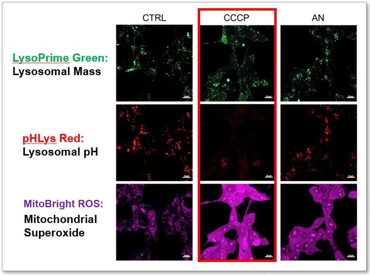
Measurement of intracellular iron changes and lysosomal pH changes
|
|||
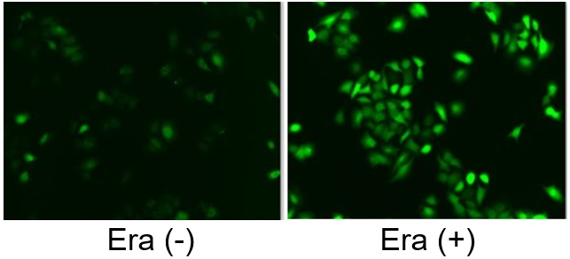
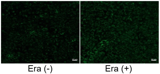
Link Lysosomal Failure to Ferroptosis in Human Neurons [May 9, 2023]
Open / Close the Article
| Here, the scientists reveal an unexpected role for the lysosomal protein prosaposin (PSAP), the knockdown of which caused the formation of lipofuscin, a hallmark of aging, which traps iron, generating reactive oxygen species and triggering ferroptosis. Intriguingly, PSAP deficiency caused these dramatic phenotypes only in neurons, but not in other cells. Learn how the authors used Dojindo's Ferroptosis-related products, FerroOrange and Liperfluo for detecting Iron levels and lipid peroxidation, in their study. | |||||
|
Genome-wide CRISPRi/a screens in human neurons link lysosomal failure to ferroptosis Ruilin Tian, et. al., Nature Neuroscience (2022) Point of Interest |
|||||
| Related Techniques | |||||
| Intracellular lipid peroxidation measurement | Liperfluo | ||||
| Mitochondria lipid peroxidation measurement | MitoPeDPP | ||||
| Intracellular ferrous ion (Fe2+) detection | FerroOrange | ||||
| Mitochondria ferrous ion (Fe2+) detection | Mito-FerroGreen | ||||
| Mitochondrial superoxide detection | MitoBright ROS Deep Red - Mitochondrial Superoxide Detection | ||||
| Lysosomal function assay | Lysosomal pH and mass detection Kit | ||||
| Cellular senescence detection (Live cell imaging or FCM) | Cellular Senescence Detection Kit | ||||
| Cellular senescence detection (Plate reader) | Cellular Senescence Plate Assay Kit | ||||
| Related Applications | |||||
The simultaneous detection of lysosomal function with Mitochondrial ROS and intracellular Fe2+
|
|||||
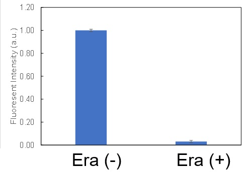
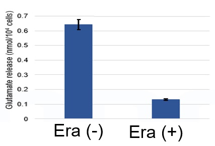
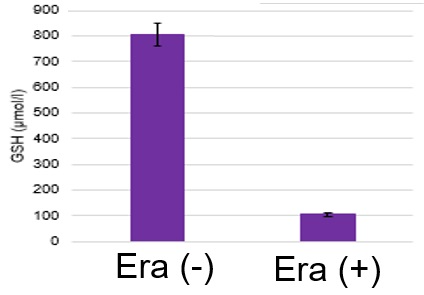
Induction of Ferroptosis by ErastinErastin is a known inducer of ferroptosis. By inhibiting the cystine transporter (xCT), erastin inhibits the uptake of cystine. Cystine is the raw material for GSH. Therefore, Erastin ultimately decreases the amount of GSH. Decreased GSH then results in lipid peroxide accumulation and induction of ferroptosis. Using erastin-treated A549 cells, we measured intracellular Fe2+, ROS, lipid peroxide, glutathione, glutamate release into the extracellular space, and cystine uptake. As a result, inhibition of xCT by elastin was observed and also the release of glutamate and uptake of cystine were decreased. Furthermore, elastin treatment decreased intracellular glutathione while it increased intracellular Fe2+ , ROS, and lipid peroxides.
|
|||||
Mechanisms and role of ferroptosis in disease [Jan. 17, 2023]
Open / Close the Article
| The field of ferroptosis research has grown exponentially in the past few years. This unique cell death by iron-dependent phospholipid peroxidation is regulated by multiple cellular metabolic pathways, including redox homeostasis, iron handling, mitochondrial activity, amino acid, lipid, and sugar metabolism, as well as various disease-related signaling pathways. Today, we introduce you to three highlighted articles focusing on iron resources, regulators, and the sensitive phenotype for ferroptosis in several diseases. | ||
|---|---|---|
| The sensitive phenotype for ferroptosis | Iron source in heart failure | Regulator of ferroptosis |
| Microglia ferroptosis is regulated by SEC24B and contributes to neurodegeneration (Sean K. Ryan, et al., Nature Neuroscience, 26, 12-26, 2023) |
Iron derived from autophagy-mediated ferritin degradation induces cardiomyocyte death and heart failure in mice (Jumpei Ito, et al., eLife, 10:e62174, 2021) |
The MARCHF6 E3 ubiquitin ligase acts as an NADPH sensor for the regulation of ferroptosis (Kha The Nguyen, et al., Nature Cell Biology, 24, 1239-1251, 2022) |
|
|
|
| Related Technique in This Topic | ||
| Intracellular lipid peroxidation measurement | Liperfluo HOT | |
| Mitochondria lipid peroxidation measurement | MitoPeDPP | |
| Mitochondria ferrous ion (Fe2+) detection | Mito-FerroGreen | |
| Intracellular ferrous ion (Fe2+) detection | FerroOrange HOT | |
| Total ROS detection | High Sensitive DCFH-DA HOT or Compatible with Immunostaining HOT | |
| Autophagy detection | DAPGreen / DAPRed (Autophagosome detection), DALGreen (Autolysosome detection) | |
| GSSG/GSH assay | GSSG/GSH Assay Kit | |
| Glutamine or Glutamate assay | Glutamine Assay Kit, Glutamate Assay Kit | |
| NADP/NADPH assay | NADP/NADPH Assay KIt | |
