


Science Note: Mitochondria
Boosting T-cells with Mitochondrial Activation to Treat Tumours [Dec. 3, 2024]
Open / Close the Article
Recent research shows that mitochondria are critical for T cell function, regulating metabolic pathways essential for immune responses. Here are some of the papers that show mechanisms and strategies, such as mitochondrial transfer or genetic modification, to improve mitochondrial health and reinvigorate T cell activity against tumours.Mitochondria play a critical role in T cell function, providing energy and regulating metabolic pathways essential for immune responses. In cancer, the tumour microenvironment induces mitochondrial dysfunction in T cells, leading to exhaustion and reduced anti-tumour efficacy. Strategies to improve mitochondrial health, such as mitochondrial transfer or genetic modification, have shown promise in reinvigorating T cell activity against tumours. Targeting mitochondrial pathways in T cells offers a novel approach to improving cancer immunotherapies and overcoming tumour-induced immunosuppression. |
|||||||||||||||
|
Intercellular nanotube-mediated mitochondrial transfer enhances T cell metabolic fitness and antitumor efficacy |
PGE2 inhibits TIL expansion by disrupting IL-2 signalling and mitochondrial function |
FOXO1 enhances CAR T cell stemness, metabolic fitness and efficacy |
|||||||||||||
|
Point of Interest - Bone marrow stromal cells transfer mitochondria to CD8+ T cells via nanotubes, requiring Talin 2 for optimal transfer. - Mitochondria-boosted T cells exhibit improved respiration, tumor infiltration, and reduced exhaustion, enhancing antitumor responses. - Intercellular mitochondrial transfer offers a new organelle-based therapy, advancing next-generation cell therapies for cancer treatment. |
Point of Interest - Prostaglandin E2 (PGE2) impairs IL-2 sensing in CD8+ tumour-infiltrating lymphocytes (TILs) by downregulating IL-2 receptor components, causing oxidative stress and cell death. - Blocking PGE2 signalling during TIL expansion restores IL-2 responsiveness, increasing TIL proliferation and improving tumour control in adoptive cell therapy. - Targeting PGE2 pathways enhances IL-2-driven T cell expansion, offering new strategies to improve cancer immunotherapy outcomes. |
Point of Interest - CAR T-cell therapy is less effective in solid tumours due to the immunosuppressive microenvironment that causes T-cell dysfunction. - Overexpression of FOXO1 enhances the stem-like phenotype, mitochondrial fitness and persistence of CAR T cells, thereby improving antitumour efficacy. - Engineering FOXO1 in CAR T cells offers a promising strategy to increase efficacy against solid tumours in cancer therapy. |
|||||||||||||
| Related Techniques | |||||||||||||||
| Mitochondrial membrane potential detection | JC-1 MitoMP Detection Kit, MT-1 MitoMP Detection Kit | ||||||||||||||
| Mitochondrial superoxide detection | MitoBright ROS Deep Red - Mitochondrial Superoxide Detection | ||||||||||||||
| Mitophagy or autophagy detection | Mitophagy Detection Kit, Autophagic Flux Assay Kit | ||||||||||||||
| Mitochondrial Staining | MitoBright LT Green / Red / Deep Red | ||||||||||||||
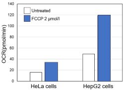
| Glycolysis/Oxidative phosphorylation Assay | Glycolysis/OXPHOS Assay Kit, Extracellular OCR Plate Assay Kit | ||||||||||||||
| Lipid Droplet Staining | Lipi-Blue/ Green/ Red/ Deep Red | ||||||||||||||
| Intracellular / mitochondrial ferrous ion (Fe2+) detection | FerroOrange, Mito-FerroGreen | ||||||||||||||
| Intracellular / mitochondrial lipid peroxidation detection | Liperfluo, MitoPeDPP | ||||||||||||||
| Apoptosis detection in multiple samples | NEW Annexin V Apoptosis Plate Assay Kit | ||||||||||||||
| Total ROS detection | Highly sensitive DCFH-DA or Photo-oxidation Resistant DCFH-DA | ||||||||||||||
| Cell Proliferation / Cytotoxicity Assay | Cell Counting Kit-8, Cytotoxicity LDH Assay Kit-WST | ||||||||||||||
| Related Applications | |||||||||||||||
|
|||||||||||||||
How mtDNA Mutations Drive Cancer Growth and New Treatments [Sep. 24, 2024]
Open / Close the Article
Recent research on cancer progression shows that mtDNA mutations affect cellular metabolism and promote tumor growth. Here are some of the studies showing how mtDNA mutations drive metabolic changes and have implications for cancer therapy.Mitochondrial DNA (mtDNA) plays a key role in cancer progression by influencing cellular metabolism and oxidative phosphorylation. Mutations in mtDNA can lead to metabolic shifts, such as the Warburg effect, that promote tumor growth and alter the tumor microenvironment. These mtDNA alterations can increase the production of reactive oxygen species (ROS), which drive pro-tumor signaling pathways such as TGFβ/Smad. In addition, mtDNA mutations and their transfer via extracellular vesicles (EVs) can affect tumor-stroma communication and potentially serve as biomarkers and therapeutic targets in cancer treatment. |
|||||||||||||
|
Mitochondrial DNA mutations drive aerobic glycolysis to enhance checkpoint blockade response in melanoma |
Mitochondrial genome transfer drives metabolic reprogramming in adjacent colonic epithelial cells promoting TGFβ1-mediated tumor progression |
Plasma cell–derived mtDAMPs activate the macrophage STING pathway, promoting myeloma progression |
|||||||||||
|
Point of Interest - DNA mutations in tumors promote metabolic shifts and anti-tumor immune responses, sensitizing them to checkpoint blockade therapies. - Engineered mtDNA mutations in melanoma reshaped the tumor microenvironment, reducing neutrophils and enhancing immune responses in mice and humans. - Tumors with >50% mtDNA mutations showed a 2.5-fold improved response to checkpoint blockade, highlighting the therapeutic potential of mtDNA. |
Point of Interest - Tumor-derived EV-mtDNA enhances mitochondrial respiration, ROS production and TGFβ1 expression, promoting colon cancer progression. - EV-mtDNA from cancer cells activates ROS-driven RelA translocation in neighboring cells, regulating TGFβ1 and promoting tumor growth. - EV-mtDNA serves as a potential biomarker and therapeutic target for paracrine metabolic communication in colon cancer. |
Point of Interest - Elevated cell-free mtDNA in multiple myeloma activates macrophages via the STING pathway and promotes disease progression in the bone marrow. - Multiple myeloma (MM) cell-derived mtDAMPs induce chemokine upregulation in bone marrow macrophages, contributing to MM cell retention and tumor growth. - Inhibition of STING signaling or chemokine response reduces multiple myeloma tumor burden and induces MM cell egress from the bone marrow. |
|||||||||||
| Related Techniques | |||||||||||||
| Glycolysis/Oxidative phosphorylation Assay | Glycolysis/OXPHOS Assay Kit, Extracellular OCR Plate Assay Kit | ||||||||||||
| Mitochondrial membrane potential detection | JC-1 MitoMP Detection Kit, MT-1 MitoMP Detection Kit | ||||||||||||
| Mitochondrial superoxide detection | MitoBright ROS Deep Red - Mitochondrial Superoxide Detection | ||||||||||||
| Mitophagy or autophagy detection | Mitophagy Detection Kit, Autophagic Flux Assay Kit | ||||||||||||
| Total ROS detection | Highly sensitive DCFH-DA or Photo-oxidation Resistant DCFH-DA | ||||||||||||
| Oxygen consumption rate assay | Extracellular OCR Plate Assay Kit | ||||||||||||
| Glutathione Quantification | GSSG/GSH Quantification Kit | ||||||||||||
| NAD(H) and NADP(H) redox couples assay | NAD/NADH and NADP/NADPH Assay Kit | ||||||||||||
| Exosome Membrane Fluorescent Staining | ExoSparkler Exosome Membrane Labeling Kit-Green/ Red/ Deep Red | ||||||||||||
| Cell Proliferation / Cytotoxicity Assay | Cell Counting Kit-8, Cytotoxicity LDH Assay Kit-WST | ||||||||||||
| Related Applications | |||||||||||||
|
|||||||||||||
Hypoxia-reprogramed mitochondria engulf lysosome for self-digestion [July 18, 2023]
Open / Close the Article
|
Scientists have unveiled that hypoxia transforms regular tubular mitochondria into larger structures called megamitochondria through inter-mitochondrial contact and fusion. During hypoxia, mitochondria-lysosome interaction is enhanced, and some lysosomes get engulfed by megamitochondria, which escalates mitochondrial ROS production. |
|||||||
|
Hypoxia-reprogramed megamitochondrion contacts and engulfs lysosome to mediate mitochondrial self-digestion Click here for the original article: Tianshu Hao, et. al., Nature Communications (2023) Point of Interest |
|||||||
| Related Techniques | |||||||
| Mitochondrial staining (Long-Term Visualization) | MitoBright LT Green, Red, Deep Red | ||||||
| Mitochondrial superoxide detection | MitoBright ROS - Mitochondrial Superoxide Detection | ||||||
| Total ROS detection | Higher sensitivity or for long-term live cell imaging | ||||||
| Lysosomal function assay | Lysosomal Acidic pH Detection kit-Green/Red, Green/Deep Red | ||||||
| Lysosomal pH Detection | pHLys Red - Lysosomal Acidic pH Detection | ||||||
| Mitochondrial membrane potential detection | JC-1 MitoMP Detection Kit / MT-1 MitoMP Detection Kit | ||||||
| Mitophagy Detection | Mitophagy Detection Kit and Mtphagy Dye | ||||||
| Oxygen consumption rate assay | Extracellular OCR Plate Assay Kit | ||||||
| Related Applications | |||||||
|
|||||||


The Role of Mitophagy in Cancer, Neuronal, and Senescent Cell Functions [Jul. 16, 2024]
Open / Close the Article
| Mitochondrial function is closely related to the onset of the neurodegenerative disorder and the aging process. From this background, much attention has been paid to elucidating the mitochondrial quality control pathway. Here, we introduce you to three individual pathways (extracellular vesicles (EVs)-related release pathway, Lysosome-related exocytosis, and Mitophagy) for mitochondrial quality control. | ||
|---|---|---|
| EVs-related release pathway | Lysosome-related exocytosis | Mitophagy |
| Ejection of damaged mitochondria and their removal by macrophages ensure efficient thermogenesis in brown adipose tissue (Marco Rosina, et al., Cell Metabolism, 34, 533-548, 2022) |
Mitolysosome exocytosis, a mitophagy-independent mitochondrial quality control in flunarizine-induced parkinsonism-like symptoms (Feixiang Bao, et al., Science Advances, 8, eabk2376, 2022) |
Neuronal induction of BNIP3-mediated mitophagy slows systemic aging in Drosophila (Edward T. Schmid, et al., Nature Aging, 2, 494-507, 2022) |
|
|
|
| Related Technique in this topic | ||
| Mitochondrial membrane potential detection | JC-1 MitoMP Detection Kit and MT-1 MitoMP Detection Kit | |
| Mitochondrial superoxide detection | MitoBright ROS - Mitochondrial Superoxide Detection | |
| Mitophagy detection | Mitophagy Detection Kit | |
| Oxygen consumption rate assay | Extracellular OCR Plate Assay Kit | |
| EVs Isolation | ExoIsolator Exosome Isolation Kit | |
| EVs labeling | ExoSparkler Exosome Membrane Labeling Kit-Green, Red, Deep red | |
| Lysosomal function assay | Lysosomal pH and mass detection Kit | |
Mitochondrial Proteins for Maintaining of Organelle Homeostasis [Jun. 11, 2024]
Open / Close the Article
|
Mitochondria are essential for maintaining lysosomal and autophagic homeostasis by providing the ATP required for lysosomal acidification and enzymatic function. They interact with lysosomes through signaling pathways and physical contacts, and coordinate the degradation and recycling of cellular components via autophagy. Efficient mitochondrial function supports the autophagic process by ensuring the removal of damaged organelles and proteins, thereby preventing cellular stress. Disruptions in mitochondrial activity can impair lysosomal function and autophagy, leading to the accumulation of cellular debris and compromised cellular homeostasis. |
||
|
The mitochondrial intermembrane space protein mitofissin drives mitochondrial fission required for mitophagy |
Mitochondrial protein C15ORF48 is a stress-independent inducer of autophagy that regulates oxidative stress and autoimmunity |
Cardiomyocyte-specific deletion of the mitochondrial transporter Abcb10 causes cardiac dysfunction via lysosomal-mediated ferroptosis |
|
Point of Interest -Mitofissin is a mitochondrial fission factor located in the mitochondrial intermembrane space that is required for receptor-mediated mitophagy. -The mechanisms and roles of membrane fission by Atg44 differ from those of the known mitochondrial fission factors, the dynamin-related proteins Dnm1. -Mitofissin-deficient cells cannot be enwrapped by the autophagosome precursor, the phagophore, due to the lack of mitochondrial fission. -Mitofissin binds directly to lipid membranes and induces lipid membrane fragility to facilitate membrane fission required for mitophagy. |
Point of Interest -Thymic epithelial cells (TECs) may use stress-independent autophagy to degrade self-protein for generation of self-antigen peptides. -The mitochondrial protein C15ORF48 induced by inflammatory stimulation reduces mitochondrial membrane potential, decreases intracellular ATP levels, activates AMPK, and ultimately induces autophagy. -Autophagy dependent on C15ORF48 increases intracellular glutathione levels and promotes cell survival by reducing oxidative stress. -C15orf48 deficient mice show a reduction in stress-independent autophagy, but not in typical starvation-induced autophagy. -C15orf48 deficient mice develop autoimmunity, and furthermore, engraftment of C15orf48-deficient thymus into nude mice results in autoimmunity. |
Point of Interest -Abcb10 is a member of the ABC transporter superfamily located in the inner mitochondrial membrane and plays an important role in iron uptake into erythrocyte mitochondria.. -Cardiomyocyte-specific Abcb10 knockout mice show progressive worsening of cardiac fibrosis, increased cardiovascular risk markers and mitochondrial structural abnormalities. -In addition, the mice exhibit increased Hif1α expression, decreased NAD synthase expression, reduced NAD+ levels and lysosomal dysfunction. -Impairment of Abcb10 leads to the accumulation of iron and lipid peroxides in lysosomes, resulting in ferroptosis and contributing to the development of chronic heart failure.
|
| Related Techniques | ||
| Mitophagy or autophagy detection | Mitophagy Detection Kit, Autophagic Flux Assay Kit | |
| Mitochondrial superoxide detection | MitoBright ROS Deep Red - Mitochondrial Superoxide Detection | |
| Mitochondrial membrane potential detection | JC-1 MitoMP Detection Kit, MT-1 MitoMP Detection Kit | |
| Lysosomal function | Lysosomal Acidic pH Detection Kit -Green/Red and Green/Deep Red | |
| Glutathione Quantification | GSSG/GSH Quantification Kit | |
| Glycolysis/Oxidative phosphorylation Assay | Glycolysis/OXPHOS Assay Kit, Extracellular OCR Plate Assay Kit | |
| Ferrous ion (Fe2+) detection | FerroOrange (intracellular), Mito-FerroGreen (mitochondrial) | |
| Lipid peroxidation detection | Liperfluo (intracellular), MitoPeDPP (mitochondrial) | |
| Cell Proliferation / Cytotoxicity Assay | Cell Counting Kit-8, Cytotoxicity LDH Assay Kit-WST | |
| Related Applications | ||
Lysosomal Function and Mitochondrial ROS
|
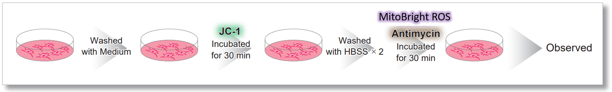
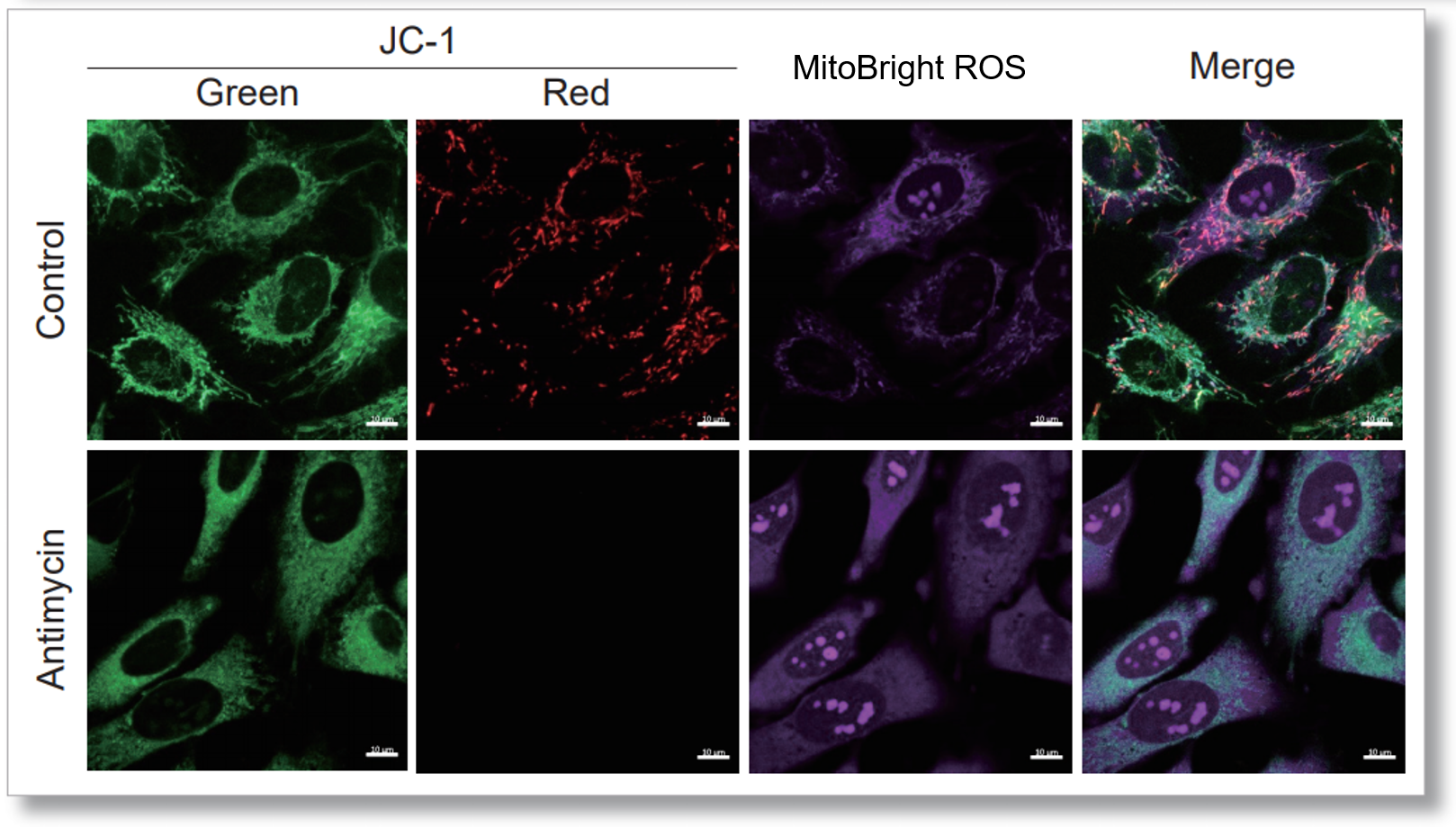
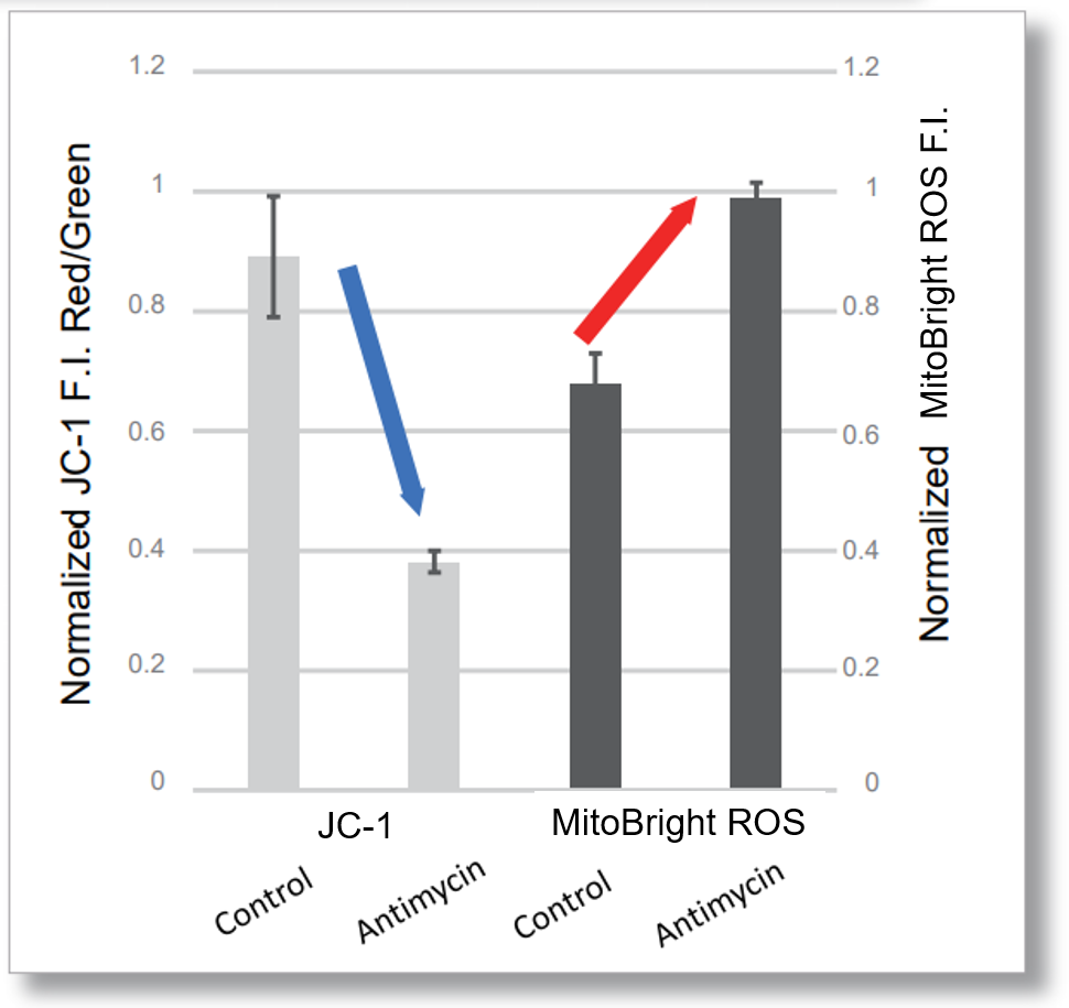
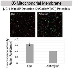
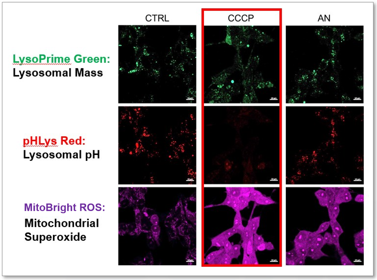
CCCP and Antimycin are recognized inducers of mitochondrial ROS, linked to the loss of mitochondrial membrane potential. Recent studies have shown that CCCP induces not only mitochondrial ROS but also lysosomal dysfunction. To observe mitochondrial ROS, HeLa cells were labeled with MitoBright ROS Deep Red for Mitochondrial Superoxide Detection, and the lysosomal mass and pH were independently detected with LysoPrime Green and pHLys Red. Co-staining with MitoBright ROS and Lysosomal dyes revealed that CCCP, unlike Antimycin, triggers concurrent lysosomal neutralization and mitochondrial ROS induction. Reference: Benjamin S Padman, et. al., Autophagy (2013) Products in Use |
|
Co-staining of Lysosome and Mitophagy
|
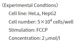
We performed fluorescence imaging by stimulating mitophagy induction in SHSY-5Y cells stained with Mitophagy Detection(Code: MD01) and LysoPrime Green or existing products. The fluorescence signal of LysoPrime Green did not decrease and the lysosomal localization over time was confirmed. This means that the co-localization rate of the fluorescent spots of the Mtphagy Dye is higher than that of the existing product, and thus more accurate mitophagy analysis can be performed.
LysoPrime Green: Ex= 488 nm, Em= 500-570 nm Products in Use |
|
Approaching Mitochondrial Cancer Therapy [May 21, 2024]
Open / Close the Article
|
Mitochondrial activity and transfer are increasingly recognized for their roles in cancer progression and therapy. Tumor cells can enhance their survival and resistance to therapy by acquiring mitochondria from surrounding stromal cells through a process known as mitochondrial transfer. This transfer not only helps cancer cells meet their increased energy and metabolic demands but also aids in evading apoptosis induced by therapeutic agents. As a result, targeting the mechanisms of mitochondrial transfer and disrupting these interactions is being explored as a novel therapeutic strategy to enhance the effectiveness of cancer treatments and reduce resistance. |
|||||||||||||
|
Mitochondrial metabolism sustains CD8+ T cell migration for an efficient infiltration into solid tumors |
Cancer cells reprogram to metastatic state through the acquisition of platelet mitochondria |
Defects of mitochondria-lysosomes communication induce secretion of mitochondria-derived vesicles and drive chemoresistance in ovarian cancer cells |
|||||||||||
|
Point of Interest - Mitochondrial oxidation of glucose and glutamine, but not fatty acids, is required for interstitial motility of CD8+ T cells. - For T cells to migrate, both ATP and ROS produced by mitochondria are required. - Increasing mitochondrial activity improves intratumoral migration of CD8+ T cells and recruitment of CAR T cells to tumor islets. |
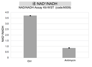
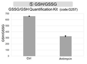
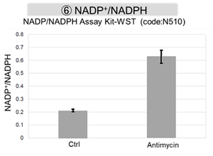
Point of Interest - Cancer cells acquire platelet mitochondria via the PINK1/Parkin-Mfn2 pathway to be reprogrammed to a metastatic state. - Transferred platelet mitochondria promoted glycolytic metabolism and increased glutathione peroxidase expression. - GSH eliminates ROS and increase, which leads to increase GSSG levels and ultimately to enhance lung metastasis of osteosarcoma in the presence of platelet mitochondria. |
Point of Interest - Cisplatin chemoresistant cells are characterized by impaired late endocytic trafficking and increased secretion of RAB7-positive mitochondria-derived vesicles (MDVs). - MDVs can be secreted by cisplatin chemoresistant cells and deliver cisplatin outside the cells. - MDVs purified from chemoresistant cells induce chemoresistance in recipient cells via a RAB7-modulated process. - The MDVs localize to mitochondria and slow down mitochondrial activity, leading to mitochondrial dysfunction, lysosomal deficit, and ultimately increased MDVs. |
|||||||||||
| Related Techniques | |||||||||||||
| Glycolysis/Oxidative phosphorylation Assay | Glycolysis/OXPHOS Assay Kit, Extracellular OCR Plate Assay Kit | ||||||||||||
| Mitochondrial membrane potential detection | JC-1 MitoMP Detection Kit, MT-1 MitoMP Detection Kit | ||||||||||||
| Mitophagy or autophagy detection | Mitophagy Detection Kit, Autophagic Flux Assay Kit | ||||||||||||
| Mitochondrial superoxide detection | MitoBright ROS Deep Red - Mitochondrial Superoxide Detection | ||||||||||||
| Total ROS detection | Highly sensitive DCFH-DA or Photo-oxidation Resistant DCFH-DA | ||||||||||||
| Lysosomal function | Lysosomal Acidic pH Detection Kit -Green/Red and Green/Deep Red | ||||||||||||
| Glutathione Quantification | GSSG/GSH Quantification Kit | ||||||||||||
| Cell Proliferation / Cytotoxicity Assay | Cell Counting Kit-8, Cytotoxicity LDH Assay Kit-WST | ||||||||||||
| Related Applications | |||||||||||||
|
|||||||||||||
Inducible and Inhibitory Organelles in Neurodegeneration [Apr. 30, 2024]
Open / Close the Article
|
In neurodegeneration, dysfunctional mitochondria contribute to oxidative stress and energy deficits in neurons. Lysosomes, responsible for the disposal of cellular waste, may be impaired, leading to the accumulation of damaged organelles, including mitochondria. At the same time, abnormal lipid metabolism and accumulation of lipid droplets have been implicated in neurodegenerative diseases, potentially exacerbating neuronal dysfunction and contributing to disease progression. The intricate relationships between dysfunctional mitochondria, impaired lysosomal function and abnormal lipid metabolism underscore the complex pathophysiology of neurodegeneration. |
|||||||||||
|
Messenger RNA transport on lysosomal vesicles maintains axonal mitochondrial homeostasis and prevents axonal degeneration |
APOE4/4 is linked to damaging lipid droplets in Alzheimer’s disease microglia |
Loss of WIPI4 in neurodegeneration causes autophagy-independent ferroptosis |
|||||||||
|
Point of Interest - Lysosome–kinesin adaptor related complex (BORC) KO depletes axonal mRNAs mainly encoding ribosomal and mitochondrial/oxidative phosphorylation proteins. - This depletion leads to mitochondrial defects and ultimately to axonal degeneration in neurons. - A mechanistic connection of BORC deficiency may accelerate common neurodegenerative disorders. |
Point of Interest - Lipid droplet-associated enzyme ACSL1-positive microglia was most abundant in patients with AD having the APOE4/4 genotype. - In microglia, fibrillar Aβ induces ACSL1 expression, triglyceride synthesis and lipid droplet accumulation depending on APOE. - Conditioned media from lipid droplet-containing microglia lead to Tau phosphorylation and neurotoxicity depending on APOE in neurons. |
Point of Interest - WIPI4 deficiency causes β-Propeller protein-associated neurodegeneration, which induces ferroptosis via an autophagy-independent mechanism in cell culture and in zebrafish. - WIPI4 depletion increases the localization of ATG2A at ER-mitochondrial contact sites, which enhances phosphatidylserine import into mitochondria. - This leads to increased mitochondrial synthesis of phosphatidylethanolamine, a major lipid prone to peroxidation and ultimately to ferroptosis. |
|||||||||
| Related Techniques | |||||||||||
| Mitochondrial or internal lipid peroxide detection | MitoPeDPP, Liperfluo | ||||||||||
| Mitochondrial or internal iron detection | Mito-FerroGreen, FerroOrange | ||||||||||
| Mitophagy or autophagy detection | Mitophagy Detection Kit, Autophagic Flux Assay Kit | ||||||||||
| Glycolysis/Oxidative phosphorylation Assay | Extracellular OCR Plate Assay Kit, Glycolysis/OXPHOS Assay Kit | ||||||||||
| Lipid droplets detection | Lipi-Blue/Green/Red/Deep Red | ||||||||||
| Lysosome staining | pH-dependent (pHLys Red) and pH resistance (LysoPrime Green/Deep Red) probes | ||||||||||
| Lysosomal acidic pH detection |
Lysosoml Acidic pH Detection Kit-Green/Red and Green/Deep Red |
||||||||||
| Related Applications | |||||||||||
|
|||||||||||
Mitochondrial Metabolism and Oxidative Stress [Feb. 6, 2024]
Open / Close the Article
|
Mitochondrial metabolism involves the biochemical processes within mitochondria that convert nutrients into energy and building blocks necessary for cell function, primarily through the citric acid cycle and oxidative phosphorylation. This energy production produces adenosine triphosphate (ATP), the cell's primary energy currency. Oxidative stress occurs when there's an imbalance between the production of reactive oxygen species (ROS) in the mitochondria and the cell's ability to detoxify these harmful byproducts or repair the resulting damage. Over time, excessive oxidative stress can lead to cellular damage that contributes to aging and several diseases, including neurodegenerative disorders and cancer. |
||||||
|
Autoregulatory control of mitochondrial glutathione homeostasis |
ApoE enhances mitochondrial metabolism via microRNA-142a/146a-regulated circuits that suppress hematopoiesis and inflammation in hyperlipidemia |
Mitochondrial protein C15ORF48 is a stress-independent inducer of autophagy that regulates oxidative stress and autoimmunity |
||||
|
Point of Interest - Depletion of GSH dissociates AFG3L2 from SLC25A39, which triggers enhancement in the uptake of GSH by mitochondria. - This regulatory mechanism is dependent on a putative iron-sulfur cluster within SLC25A39, linking the control of mitochondrial iron homeostasis to GSH import. |
Point of Interest - By diminishing miR-142a levels, ApoE enhances fatty acid oxidation, thereby improving mitochondrial metabolism. - ApoE plays a critical role in regulating immune cell metabolism, influencing hematopoiesis and inflammation in conditions of hyperlipidemia. |
Point of Interest - C15ORF48-dependent induction of autophagy upregulates intracellular glutathione levels and promotes cell survival by reducing oxidative stress. - Mice deficient in C15orf48 show a reduction in stress-independent autophagy in thymic epithelial cells (TECs). - C15orf48-/- mice develop autoimmunity, suggesting that stress-independent autophagy in TECs is critical for thymic self-tolerance. |
||||
| Related Techniques | ||||||
| Oxygen consumption rate assay | Extracellular OCR Plate Assay Kit | |||||
| Mitochondrial superoxide detection | MitoBright ROS - Mitochondrial Superoxide Detection NEW | |||||
| Mitophagy Detection | Mitophagy Detection Kit and Mtphagy Dye | |||||
| Mitochondrial membrane potential detection | JC-1 MitoMP Detection Kit / MT-1 MitoMP Detection Kit | |||||
| Glycolytic/Mitochondrial activity detection | Glycolysis/JC-1 MitoMP Assay Kit NEW | |||||
| Glycolysis/Oxidative phosphorylation Assay | Glycolysis/OXPHOS Assay Kit | |||||
| Mitochondrial staining (Long-Term Visualization) | MitoBright LT Green, Red, Deep Red | |||||
| Total ROS detection | Highly sensitive DCFH-DA or Photo-oxidation Resistant DCFH-DA | |||||
| Antibody/Protein labeling with fast and high recovery |
Fluorescein, Biotin, and Peroxidase Labeling Kit - NH2 | |||||
| Related Applications | ||||||
|
||||||
Mitochondrial Stress Responses [Jan. 9, 2024]
Open / Close the Article
|
Mitochondrial stress responses are a set of cellular reactions triggered when mitochondria, the energy-producing organelles in cells, encounter various forms of stress. This can include damage from reactive oxygen species (ROS), fluctuations in nutrient levels, or other environmental stressors. In response to such stress, mitochondria can undergo changes in their dynamics, such as fission and fusion, to maintain cellular homeostasis and energy production. Additionally, cells activate protective pathways, like the mitochondrial unfolded protein response (UPRmt), to repair or degrade damaged mitochondrial proteins and mitigate the potentially harmful effects of mitochondrial dysfunction. |
||||||||||||
|
Mitochondrial fission drives neuronal metabolic burden to promote stress susceptibility in male mice |
Mitochondrial DNA breaks activate an integrated stress response to reestablish homeostasis |
Glial-derived mitochondrial signals affect neuronal proteostasis and aging |
||||||||||
|
Point of Interest - Enhancing Drp1-dependent mitochondrial fission promotes stress susceptibility. - Increased stress susceptibility is alleviated by increasing mitochondrial ATP production. |
Point of Interest - mtDSBs initiate an integrated stress response (ISR). - Inhibition of the ISR exacerbates mitochondrial defects and slows recovery of mitochondrial DNA. - ATAD3A is a potential signaling molecule from damaged genomes to the inner membrane of mitochondria. |
Point of Interest - Glial cells use small clear vesicles (SCVs) to signal the mitochondrial proteotoxic stress to neurons. - Neurons relay the signal to the periphery using dense-core vesicles. |
||||||||||
| Related Techniques | ||||||||||||
| Oxygen consumption rate assay | Extracellular OCR Plate Assay Kit | |||||||||||
| Mitochondrial superoxide detection | MitoBright ROS - Mitochondrial Superoxide Detection | |||||||||||
| Total ROS detection | Highly sensitive DCFH-DA or Photo-oxidation Resistant DCFH-DA | |||||||||||
| Mitophagy Detection | Mitophagy Detection Kit and Mtphagy Dye | |||||||||||
| Mitochondrial membrane potential detection | JC-1 MitoMP Detection Kit / MT-1 MitoMP Detection Kit | |||||||||||
| Glycolytic/Mitochondrial activity detection | Glycolysis/JC-1 MitoMP Assay Kit | |||||||||||
| Glycolysis/Oxidative phosphorylation Assay | Glycolysis/OXPHOS Assay Kit | |||||||||||
| Mitochondrial staining (Long-Term Visualization) | MitoBright LT Green, Red, Deep Red | |||||||||||
| Antibody/Protein labeling with fast and high recovery | Fluorescein, Biotin, and Peroxidase Labeling Kit - NH2 | |||||||||||
| Related Applications | ||||||||||||
|
||||||||||||
Mitochondrial Stress Responses: Iron, ROS, and Mitophagy [Aug 1, 2023]
Open / Close the Article
|
In recent years, the discovery of several novel mitochondrial stress responses has attracted much attention. A mitochondrial iron-responsive pathway was discovered that protects cells from iron deficiency. In other breakthroughs, a new anticancer target is identified and the nucleus-to-mitochondria ROS-sensing pathway is implicated in resistance to platinum-based treatments for ovarian cancer. Research also uncovers the role of the protein in promoting mitochondrial fission, essential for effective mitophagy. |
||||||||||||
|
A mitochondrial iron-responsive pathway regulated by DELE1 |
Systematic identification of anticancer drug targets reveals a nucleus-to-mitochondria ROS-sensing pathway |
The mitochondrial intermembrane space protein mitofissin drives mitochondrial fission required for mitophagy |
||||||||||
|
Point of Interest |
Point of Interest |
Point of Interest |
||||||||||
| Related Techniques | ||||||||||||
| Mitochondria ferrous ion (Fe2+) detection | Mito-FerroGreen | |||||||||||
| Mitochondrial superoxide detection | MitoBright ROS - Mitochondrial Superoxide Detection | |||||||||||
| Mitophagy Detection | Mitophagy Detection Kit and Mtphagy Dye | |||||||||||
| Mitochondrial membrane potential detection | JC-1 MitoMP Detection Kit / MT-1 MitoMP Detection Kit | |||||||||||
| Glycolytic/Mitochondrial activity detection | Glycolysis/JC-1 MitoMP Assay Kit | |||||||||||
| Oxygen consumption rate assay | Extracellular OCR Plate Assay Kit | |||||||||||
| Mitochondrial staining (Long-Term Visualization) | MitoBright LT Green, Red, Deep Red | |||||||||||
| Total ROS detection | Higher sensitivity or for long-term live cell imaging | |||||||||||
| Related Applications | ||||||||||||
|
||||||||||||
Iron-Related Autophagy and Mitochondrial Homeostasis [May 31, 2023]
Open / Close the Article
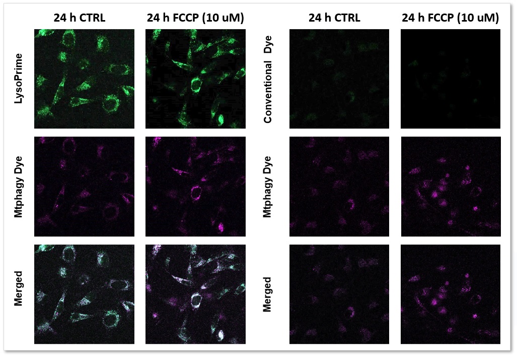
| Scientists have unveiled that the selective autophagy adaptor NCOA4, which targets ferritin (a process known as ferritinophagy), is upregulated in pancreatic cancer. This upregulation ensures the availability of iron, thus encouraging tumor progression. Fascinatingly, ferritinophagy facilitates the synthesis of iron–sulfur cluster proteins, which help maintain mitochondrial homeostasis. Discover how the authors employed separate iron-detecting probes: FerroOrange for the cytosol and Mito-FerroGreen for the mitochondria (refer to Figure 1I and Supplemental Figure S4J). | |||||
|
NCOA4-Mediated Ferritinophagy Is a Pancreatic Cancer Dependency via Maintenance of Iron Bioavailability for Iron–Sulfur Cluster Proteins Naiara Santana-Coodina, et. al., Cancer Discovery (2022) Point of Interest |
|||||
| Related Techniques | |||||
| Intracellular ferrous ion (Fe2+) detection | FerroOrange | ||||
| Mitochondria ferrous ion (Fe2+) detection | Mito-FerroGreen | ||||
| Mitophagy Detection | Mitophagy Detection Kit | ||||
| Lysosomal function assay | Lysosomal pH and mass detection Kit | ||||
| Mitophagy Detection | Mitophagy Detection Kit and Mtphagy Dye | ||||
| Autophagy detection | DAPGreen / DAPRed (Autophagosome detection), DALGreen (Autolysosome detection) | ||||
| Oxygen consumption rate assay | Extracellular OCR Plate Assay Kit | ||||
| Related Applications | |||||
The simultaneous detection of lysosomal function with Mitochondrial ROS and intracellular Fe2+
|
|||||
Mitochondrial unfolded protein response in neural stem cell aging [May 9, 2023]
Open / Close the Article
| This article focusing on the mitochondrial protein folding stress is particularly significant in activated neural stem cells (NSCs) and neural progenitor cells (NPCs) within the neurogenic niche. This stress increases with age, leading to dysregulated cell cycles and mitochondrial activity in activated NSCs and NPCs in the dentate gyrus. | |||||
|
The mitochondrial unfolded protein response regulates hippocampal neural stem cell aging Chin-Ling Wang, et. al., Cell Metabolism (2023) Point of Interest |
|||||
| Related Techniques | |||||
| Glycolysis/Oxidative phosphorylation assay | Glycolysis/OXPHOS Assay Kit | ||||
| Oxygen consumption rate assay | Extracellular OCR Plate Assay Kit | ||||
| Mitochondrial membrane potential detection | JC-1 MitoMP Detection Kit / MT-1 MitoMP Detection Kit | ||||
| Mitochondrial superoxide detection | MitoBright ROS - Mitochondrial Superoxide Detection | ||||
| Mitophagy Detection | Mitophagy Detection Kit and Mtphagy Dye | ||||
| Cellular senescence detection (Live cell imaging or FCM) | Cellular Senescence Detection Kit | ||||
| Cellular senescence detection (Plate reader) | Cellular Senescence Plate Assay Kit | ||||
| Lysosomal function assay | Lysosomal pH and mass detection Kit | ||||
| Related Applications | |||||
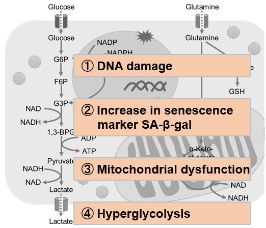
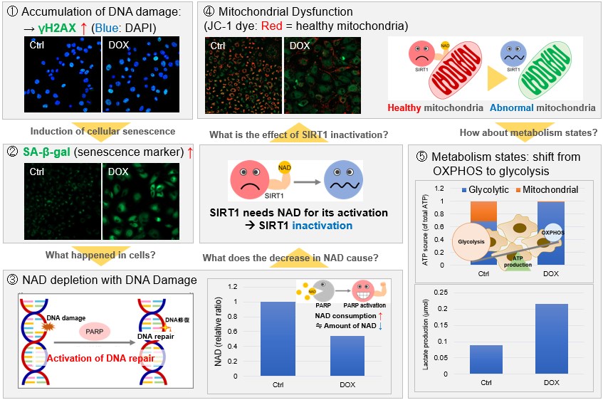
Metabolic shift to glycolysis in senescenct cells
|
|||||
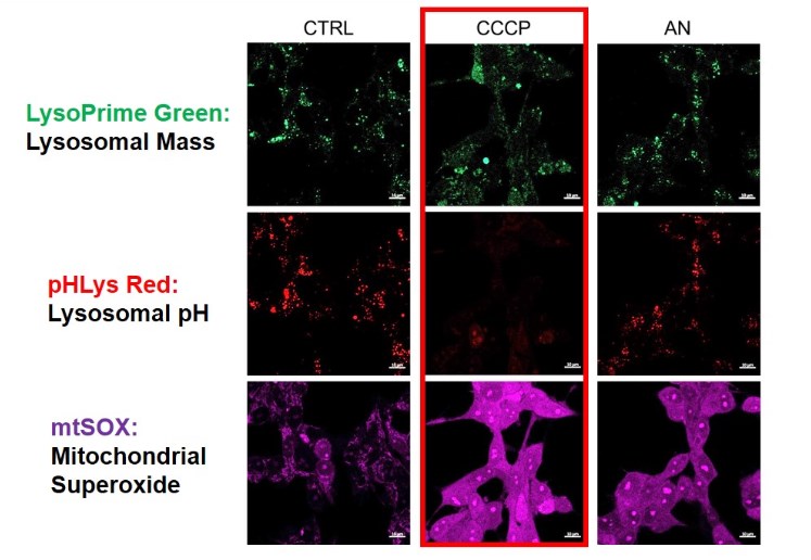
2. Simultaneously detection of Lysosomal and Mitochondrial Dysfunction
We tried the simultaneous detection of lysosomal and mitochondrial dysfunction in Hela cells treated with CCCP or Antimycin (AN). CCCP and AN are well-known inducers of mitochondrial ROS regarding loss of mitochondrial membrane potential. Recent research showed the result that CCCP induces not only mitochondrial ROS but also lysosomal neutralization. To detect mitochondrial ROS, HeLa cells were labeled by MitoBright ROS - Mitochondrial Superoxide Detection, and the lysosomal mass and pH were detected separately with LysoPrime Green and pHLys Red. Co-staining with MitoBright ROS and Lysosomal dyes demonstrated that CCCP causes lysosomal neutralization and mitochondrial ROS induction at the same time. Products in Use
|
|||||
Macrophage Necrosis through a Mitochondrial-Lysosomal-ER Circuit [May 2, 2023]
Open / Close the Article
| This article focusing on the mechanisms of programmed necrosis in infected macrophages. The research demonstrates that the excess tumor necrosis factor triggers programmed necrosis of infected macrophages is not mitochondrion-intrinsic but results from an inter-organellar circuit initiating and culminating in the mitochondrion. The circuit begins and ends with the transit of two inorganic signals - ROS from mitochondrion to lysosome and Ca2+ from endoplasmic reticulum to mitochondrion - and requires cathepsin D translocation from lysosome to cytosol. | |
|
TNF Induces Pathogenic Programmed Macrophage Necrosis in Tuberculosis through a Mitochondrial-Lysosomal-Endoplasmic Reticulum Circuit Francisco J. Roca, et. al., Cell (2019) Point of Interest |
|
| Related Techniques | |
| Total ROS detection | Higher sensitivity or for long-term live cell imaging HOT |
| Mitochondrial superoxide detection | MitoBright ROS - Mitochondrial Superoxide Detection |
| Glycolysis/Oxidative phosphorylation Assay | Glycolysis/OXPHOS Assay Kit |
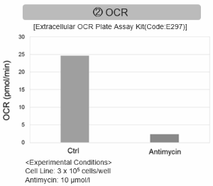
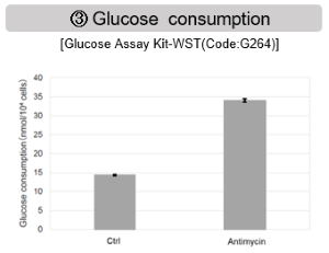
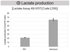
| Glycolysis-related metabolites assay | Glucose and Lactate Assay Kit |
| Oxygen consumption rate assay | Extracellular OCR Plate Assay Kit |
| Mitochondrial membrane potential detection | JC-1 MitoMP Detection Kit / MT-1 MitoMP Detection Kit |
| Mitophagy Detection | Mitophagy Detection Kit and Mtphagy Dye |
| Lysosomal function assay | Lysosomal pH and mass detection Kit |
| Autophagy detection | DAPGreen / DAPRed (Autophagosome detection), DALGreen (Autolysosome detection) |
| Related Applications | |
1. Simultaneously detection of Lysosomal and Mitochondrial Dysfunction
We tried the simultaneous detection of lysosomal and mitochondrial dysfunction in Hela cells treated with CCCP or Antimycin (AN). CCCP and AN are well-known inducers of mitochondrial ROS regarding loss of mitochondrial membrane potential. Recent research showed the result that CCCP induces not only mitochondrial ROS but also lysosomal neutralization. To detect mitochondrial ROS, HeLa cells were labeled by MitoBright ROS - Mitochondrial Superoxide Detection, and the lysosomal mass and pH were detected separately with LysoPrime Green and pHLys Red. Co-staining with mitoBright ROS and Lysosomal dyes demonstrated that CCCP causes lysosomal neutralization and mitochondrial ROS induction at the same time. Products in Use
|
|
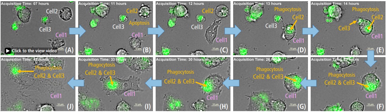
2. Monitoring ROS in Macrophage PhagocytosisDead cells (2&3) phagocytosed by Cell1 resulted in increased ROS(green). ROS detection reagent allowed for reliable analysis of the role of ROS in phagocytosis. Its high intracellular residence and low background noise made it possible to perform long-term analysis of ROS production in the cell. This information can provide important insights into the mechanisms of phagocytosis and contribute to the development of treatments for diseases associated with macrophage dysfunction. > for detail experimental notes are available at Nikon web site. Products in Use
|
|
Lipid Homeostasis Controlled by Astrocytic OxPhos [Apr. 25, 2023]
Open / Close the Article
| This article focusing on an intriguing study that emphasizes the critical role of astrocytic mitochondrial dysfunction in neurodegeneration and neuroinflammation, closely resembling Alzheimer's disease. The study underscores the significance of lipid homeostasis controlled by astrocytic OxPhos in maintaining brain health. We hope you find this information informative and inspiring for your research endeavors. | |
|
Loss of fatty acid degradation by astrocytic mitochondria triggers neuroinflammation and neurodegeneration Point of Interest |
|
| Related Techniques | |
| Total ROS detection | ROS Assay Kit -Highly Sensitive DCFH-DA- HOT |
| ROS Assay Kit -Photo-oxidation Resistant DCFH-DA- HOT | |
| Mitochondrial superoxide detection | MitoBright ROS - Mitochondrial Superoxide Detection |
| Glycolysis/Oxidative phosphorylation Assay | Glycolysis/OXPHOS Assay Kit |
| Glycolysis-related metabolites assay | Glucose and Lactate Assay Kit |
| Oxygen consumption rate assay | Extracellular OCR Plate Assay Kit |
| Mitochondrial membrane potential detection | JC-1 MitoMP Detection Kit / MT-1 MitoMP Detection Kit |
| Intracellular lipid peroxidation measurement | Liperfluo |
| Lipid droplets detection and fatty acid uptake | Lipi-Blue / Green / Red / Deep Red, Fatty Acid Uptake Assay Kit |
| Related Applications | |
1. Monitoring ROS in Macrophage PhagocytosisDead cells (2&3) phagocytosed by Cell1 resulted in increased ROS(green). ROS detection reagent allowed for reliable analysis of the role of ROS in phagocytosis. Its high intracellular residence and low background noise made it possible to perform long-term analysis of ROS production in the cell. This information can provide important insights into the mechanisms of phagocytosis and contribute to the development of treatments for diseases associated with macrophage dysfunction. > for detail experimental notes are available at Nikon web site. Products in Use
|
|
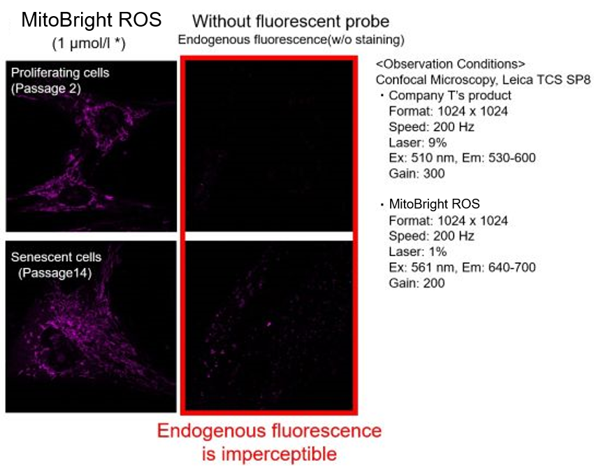
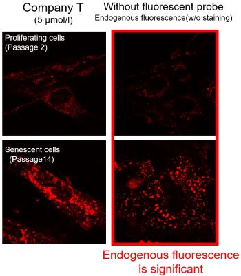
2. Mitochondrial Superoxide Detection in Senescent CellsBackground fluorescence caused by lipofuscin can be minimized by using a better fluorescent probe, as tested in TIG-1 cells. Products in Use
|
|

Mitohormesis and Inflammation [Apr. 18, 2023]
Open / Close the Article
| This article focusing on how both drug-induced and lipopolysaccharide (LPS)-induced mitochondrial stress in macrophages triggers a stress response called mitohormesis. Mitohormesis serves as a negative feedback mechanism to restrain inflammation. These findings have the potential to contribute to the development of novel strategies for counteracting acute and chronic inflammation by leveraging the Mitochondria-ROS stress response. | |
|
Mitohormesis reprogrammes macrophage metabolism to enforce tolerance Point of Interest |
|
| Related Techniques | |
| Total ROS detection | ROS Assay Kit -Highly Sensitive DCFH-DA- HOT |
| ROS Assay Kit -Photo-oxidation Resistant DCFH-DA- HOT | |
| Mitochondrial superoxide detection | MitoBright ROS - Mitochondrial Superoxide Detection |
| Glycolysis-related metabolites assay | Glucose and Lactate Assay Kit |
| Oxygen consumption rate assay | Extracellular OCR Plate Assay Kit |
| Mitochondrial membrane potential detection | JC-1 MitoMP Detection Kit / MT-1 MitoMP Detection Kit |
| Lysosomal function assay | Lysosomal pH and mass detection Kit HOT |
| Autophagy detection | DAPGreen / DAPRed (Autophagosome detection), DALGreen (Autolysosome detection) |
Conventional and Unique Quality Control Pathway of Mitochondria [Jan. 10, 2023]
Open / Close the Article
| Mitochondrial function is closely related to the onset of the neurodegenerative disorder and the aging process. From this background, much attention has been paid to elucidating the mitochondrial quality control pathway. Here, we introduce you to three individual pathways (extracellular vesicles (EVs)-related release pathway, Lysosome-related exocytosis, and Mitophagy) for mitochondrial quality control. | ||
|---|---|---|
| EVs-related release pathway | Lysosome-related exocytosis | Mitophagy |
| Ejection of damaged mitochondria and their removal by macrophages ensure efficient thermogenesis in brown adipose tissue (Marco Rosina, et al., Cell Metabolism, 34, 533-548, 2022) |
Mitolysosome exocytosis, a mitophagy-independent mitochondrial quality control in flunarizine-induced parkinsonism-like symptoms (Feixiang Bao, et al., Science Advances, 8, eabk2376, 2022) |
Neuronal induction of BNIP3-mediated mitophagy slows systemic aging in Drosophila (Edward T. Schmid, et al., Nature Aging, 2, 494-507, 2022) |
|
|
|
| Related Technique in this topic | ||
| Mitochondrial membrane potential detection | JC-1 MitoMP Detection Kit and MT-1 MitoMP Detection Kit | |
| Mitochondrial superoxide detection | MitoBright ROS - Mitochondrial Superoxide Detection | |
| Mitophagy detection | Mitophagy Detection Kit | |
| Oxygen consumption rate assay | Extracellular OCR Plate Assay Kit | |
| EVs Isolation | ExoIsolator Exosome Isolation Kit | |
| EVs labeling | ExoSparkler Exosome Membrane Labeling Kit-Green, Red, Deep red | |
| Lysosomal function assay | Lysosomal pH and mass detection Kit | |
