-Cellstain- DAPI
Dead Cell Staining
-
Product codeD212 -Cellstain- DAPI
-
CAS No.28718-90-3
-
Chemical name4',6-Diamidino-2-phenylindole, dihydrOchloride
-
MWC16H17Cl2N5=350.25
| Unit size | Price | Item Code |
|---|---|---|
| 1 mg | $79.00 | D212-10 |
Product Description
DAPI is an AT-sequence-specific DNA intercalator that attaches to DNA at the minor groove of the double helix like Hoechst dyes. DAPI is not permeable through viable cell membranes, but it passes through disturbed cell membranes to stain the nucleus. DAPI has a high photobleaching tolerance level. DAPI is utilized for the detection of mitochondrial DNA in yeast, chloroplast DNA, virus DNA, micoplasm DNA, and chromosomal DNA. The excitation and emission wavelengths of DAPI-DNA complex are 360 nm and 460 nm, respectively.
Chemical Structure


Technical info
Staining Procedure
1.Prepare 10-50 μM DAPI solution with PBS or an appropriate buffer.a)
2.Add DAPI solution with 1/10 of the volume of cell culture medium to the cell culture.b)
3.Incubate the cell at 37oC for 10-20 min.
4.Wash cells twice with PBS or an appropriate buffer.
5.Observe the cells using a fluorescence microscope with 360 nm excitation and 460 nm emission filters.
a) Since DAPI may be carcinogenic, extreme care is necessary during handling.
b) Or you may replace the culture medium with 1/10 concentration of DAPI buffer solution.
Staining Data
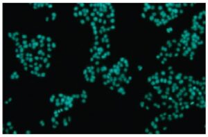
Fig. 2 Cell staining with DAPI
References
1. W. Schnedl, et al., DIPI and DAPI: Fluorescence Banding with Only Negligible Fading. Hum Genet. 1977;36:167-172.
2. I. W. Taylor, et al., An Evaluation of DNA Fluochromes, Staining Techniques, and Analysis for Flow Cytometry. I. Unperturebed Cellpopulations. J Histochem Cytochem. 1980;28:1224-1232.
3. F. Otto, et al., A Comparative Study of DAPI, DIPI, and Hoechst 33258 and 33342 as Chromosomal DNA Stains. Stain Technol. 1985;60:7-11.
4. N. Poulin, et al., Quantitative Precision of an Automated Image Cytometric System for the Measurement of DNA Content and Distribution in Cells Labeled with Fluorescent Nucleic Acid Stains. Cytometry. 1994;16:227-235.
5. M. Kawai, et al., Rapid Enumeration of Physiologically Active Bacteria in Purified Water Used in the Pharmaceutical Manufacuturing Process. J Appl Microbiol. 1999;86:496-504.
Handling and storage condition
| Appearance: | Yellow to yellow greenish brown powder or solid |
|---|---|
| Solubility in water: | To pass test (clear, pale yellow) |
| NMR spectrum: | Authentic |
| -20°C, Protect from light |
Related products
The following related products are also used in research related to this product.