Ab-10 Rapid Biotin Labeling Kit

Antibody Labeling
- Small Quantity of Antibody Requirement : 10 ug Antibody Sample Labeling
- Easy Labeling Procedure : Just Mix Antibody and Labeling Agent
- Rapid Labeling : Less than 30 minutes
-
Product codeLK37 Ab-10 Rapid Biotin Labeling Kit
| Unit size | Price | Item Code |
|---|---|---|
| 3 samples | $247.00 | LK37-10 |
| 3 samples | ・Reactive Biotin ・Reaction Buffer ・Stop Solution |
x3 100 μlx1 100 μlx1 |
|---|
Description
Product Description
Ab-10 Rapid Biotin Labeling Kit enables rapid (in less than 30 min) and easy labeling of Biotin to 10 μg antibody. Reactive Biotin (a component of the kit) has succinimidyl ester group, that can easily make a covalent bond with an amino group of the target antibody without any activation process. This kit contains all the necessary reagents to prepare a Biotin-labeled antibody except for DMSO.

| Developer | Dojindo Molecular Technologies, Inc. |
|---|
Manual
Technical info
♦ Use 0.5-1 mg/ml of antibody solution for labeling. If the antibody concentration is more than 1 mg/ml, please dilute the antibody solution with PBS.
♦ If the sample solution contains small insoluble materials, centrifuge the solution, and use the supernatant for the labeling.
♦ The microtubes in this kit contain solutions. Since there is a possibility that the droplets might attach to the inside walls or caps, please spin the tube to drop them down prior to open.
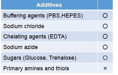
♦ In case an antibody solution includes a high concentration of constituents, such as BSA or glycerol, it may interfere with a labeling and cause a non-specific signal. We recommend removing the constituents prior to labeling procedure. Usable constituents (○) and non-usable constituents (×) are shown in Table 1, and compatible concentrations of constituents are shown in Table 2.
♦ Interference and non-specific signal may be dependent on types of antigen, host species of antibody or constituents.
♦ Since reactive reagent binds to an amino group in antibody, there is a possibility that the labeled antibody loses the antigen recognition ability (Table 3).
Table. 1 Usable / Non-usable Constituents

Table. 2 Compatible Concentration of Constituents

Table. 3 Non-Compatible Antibody

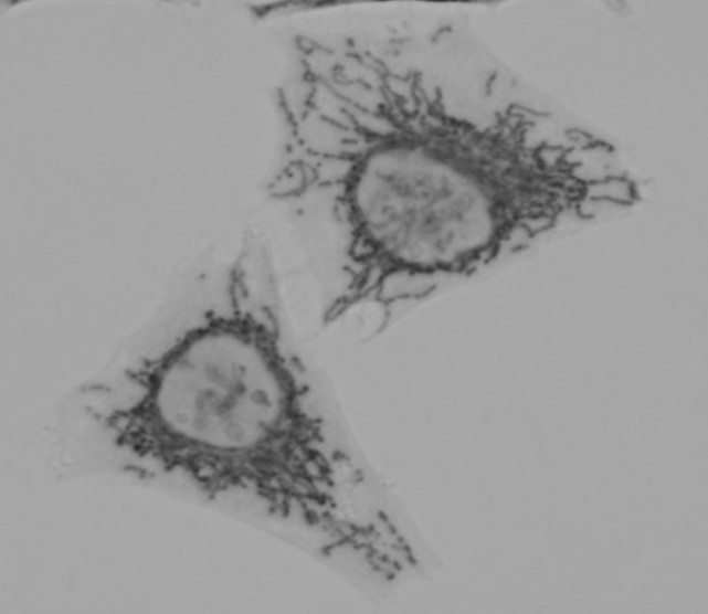
Mitochondria Immunostaining
1. HeLa cells were seeded on a μ-slide 8 well (ibidi) and cultured overnight at 37 ºC in a 5% CO2 incubator.
2. The cells were washed with PBS three times, and 4% paraformaldehyde in PBS was added to the μ-slide.
3. The μ-slide was incubated at room temperature for 15 minutes.
4. The cells were washed with PBS three times, and 1% Triton-X in PBS was added to the μ-slide.
5. The μ-slide was incubated at room temperature for 30 minutes.
6. Once the cells were washed with PBS three times, a blocking solution prepared with PBS was added to the μ-slide.
7. The cells were then incubated at room temperature for 1 hour.
8. Biotin conjugated anti-mitochondria antibody was diluted 50 times with the blocking solution. ※Anti-mitochondria antibody was purchased from Abcam (Product Code: ab3298) .
9. The supernatant was discarded and the solution (step 8) was added to the μ-slide.
10. The μ-slide was incubated at 0-5ºC overnight.
11. The supernatant was discarded and the cells were washed using PBS-T three times.
12. 0.2 µg/ml peroxidase conjugated streptavidin was added to the μ-slide.
13. The μ-slide was incubated at room temperature for 1 hour.
14. The supernatant was discarded and the cells were washed using PBS-T three times.
15. The cells were washed using Tris buffer (TB, 50 mmol/l, pH 7.5) three times.
16. The supernatant was discarded and DAB solution [0.2 mg/ml DAB (SKU:D006), 0.003% H2O2 , 50 mmol/l Tris (pH 7.5)] was added to the μ-slide.
17. The μ-slide was incubated at room temperature for 10 minutes.
18. After the cells were washed using TB three times, TB was added to the μ-slide.
19.The cells were observed under a microscope.

Fig. 2 Microscope image of DAB-stained mitochondria in HeLa cells

Handling and storage condition
| 0-5°C, Protect from moisture |


