-Cellstain- PI solution

Dead Cell Staining
-
Product codeP378 -Cellstain- PI solution
-
CAS No.25535-16-4(PI)
-
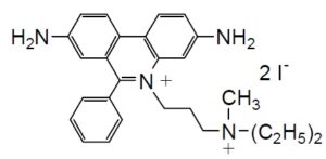
Chemical name3,8-Diamino-5-[3-(diethylmethylammonio)propyl]-6-phenylphenanthridinium diiodide, solution
-
MWC27H34I2N4=668.39
| Unit size | Price | Item Code |
|---|---|---|
| 1 ml | $93.00 | P378-10 |
Description
Product Description
Propidium iodide (PI) is an ethidium bromide analog that emits red fluorescence upon intercalation with double-stranded DNA. PI does not permeate viable cell membranes, but passes through disturbed cell membranes and stains the nuclei. PI is often used in combination with a fluorescein compound, such as Calcein-AM or FDA, for simultaneous staining of viable and dead cells. The excitation and emission wavelengths of PI-DNA complex are 535 nm and 615 nm, respectively.
Chemical Structure

Technical info
1.Prepare 10-50 μM PI solution with PBS or an appropriate buffer.a)
2.Add PI solution with 1/10 of the volume of cell culture medium to the cell culture.b)
3.Incubate the cell at 37oC for 10-20 min.
4.Wash cells twice with PBS or an appropriate buffer.
5.Observe the cells under a fluorescence microscope with 535 nm excitation and 615 nm emission filters.
a) Since PI may be carcinogenic, extreme care is necessary during handling. b) Or you may replac
e the culture medium with 1/10 concentration of PI buffer solution.
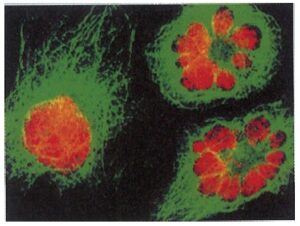
Staining Data

Fig. 2 Cell staining with PI
References
1) I. W. Taylor and B. K. Milthorpe, "An Evaluation of DNA Fluochromes, Staining Techniques, and Analysis for Flow Cytometry. I. Unperturebed Cellpopulations", J. Histochem. Cytochem., 1980, 28(11), 1224.
2) W. M. J. Vuist, F. V. Buitenen, M. A. De Rie, A. Hekman, P. Ruemke and C. J. M. Melief, "Potentiation by Interleukin 2 of Burkitt's Lymphoma Therapy with Anti-Pan B (Anti-CD19) Monoclonal Antibodies in a Mouse Xenotransplantation Model", Cancer Res., 1989, 49, 3783.
3) A. K. El-Naggar, J. G. Batsakis, K. Teague, L. Garnsey and B. Barlogie, "Single- and Double-stranded RNA Measurements by Flow Cytometry in Solid Neoplasms", Cytometry, 1991, 12, 330.
4) C. Souchier, M. Ffrench, M. Benchaib, R. Catallo and P. A. Bryon, "Methods for Cell Proloferation Analysis by Fluorescent Image Cytometry", Cytometry, 1995, 20, 203.
5)T. Yamazaki, H. Suzuki, S. Yamada, K. Ohshio, M. Sugamata, T. Yamada and Y. Morita, "Lactobacillus paracasei KW3110 Suppresses Inflammatory Stress-Induced Premature Cellular Senescence of Human Retinal Pigment Epithelium Cells and Reduces Ocular Disorders in Healthy Humans”, Int J Mol Sci, 2020, 21(14), 5091
Handling and storage condition
| Appearance: | Orange to red liquid |
|---|---|
| Dye content: | To pass test |
| -20°C, Protect from light |


