|
Recent research shows that lysosomal dysfunction in cells leads to toxic accumulation, causing stress responses, cell senescence and cell death. Here are some of the papers showing that lysosomal homeostasis is important for disease prevention.
Lysosomal dysfunction disrupts essential cellular processes and leads to the promotion of several diseases, including neurodegenerative diseases and senescence. In neurodegenerative diseases such as Alzheimer's and Parkinson's, impaired lysosomal activity leads to the accumulation of toxic proteins that contribute to neuronal death and disease progression. Lysosomal dysfunction is also associated with cellular senescence, where it impairs the degradation of damaged cellular components, leading to the accumulation of senescent cells and promoting age-related diseases. Therapeutic strategies targeting lysosomal function are being explored to alleviate these conditions and improve overall cellular health.
|
|
-
PLD3 and PLD4 synthesize S,S-BMP, a key phospholipid enabling lipid degradation in lysosomes
Click here for the original article: Shubham Singh, et. al., Cell, 2024.
Point of Interest
- PLD3 and PLD4 synthesize lysosomal S,S-bis(monoacylglycero)phosphate (BMP), which is critical for lipid degradation and brain health.
- Lysosomal S,S-BMP is essential for lipid degradation; its synthesis by PLD3/4 prevents gangliosidosis and supports cellular health.
- Mutations in PLD3 associated with neurodegenerative diseases reduce its activity, affecting S,S-BMP levels and contributing to lysosomal dysfunction.
-
Modeling Parkinson’s disease pathology in human dopaminergic neurons by sequential exposure to α-synuclein fibrils and proinflammatory cytokines
Click here for the original article: Armin Bayati, et. al., Nature Neuroscience, 2024.
Point of Interest
- Immune-induced lysosomal dysfunction promotes Lewy body (LB) formation in dopaminergic neurons, mimicking Parkinson's disease (PD) pathology.
- α-Synuclein fibrils and immune challenges (e.g., interferon-γ treatment) induce LD-like inclusions in iPSC-derived dopaminergic neurons.
- LB formation in PD may result from lysosomal-autophagic dysfunction, exacerbated by immune challenge and α-synuclein accumulation.
-
HKDC1, a target of TFEB, is essential to maintain both mitochondrial and lysosomal homeostasis, preventing cellular senescence
Click here for the original article: Mengying Cui, et. al., PNAS, 2024.
Point of Interest
- Mitochondrial and lysosomal functions are linked, with transcription factor EB (TFEB) and hexokinase domain containing 1 (HKDC1) playing critical roles in their regulation and maintenance.
- HKDC1, regulated by TFEB, is essential for mitophagy and lysosomal repair, independent of its glycolysis function.
- Loss of HKDC1 accelerates cellular senescence by causing mitochondrial hyperfusion and lysosomal damage.
|
|
Related Techniques
|
- Lysosomal function
- Lysosomal Acidic pH Detection Kit -Green/Red and Green/Deep Red
|
- Mitophagy detection
- Mitophagy Detection Kit
|
- First-time autophagy research
- Autophagic Flux Assay Kit
|
- Autophagy detection
- DAPGreen/ DAPRed (Autophagosome detection), DALGreen (Autolysosome detection)
|
|
|
- Cellular senescence detection
-
|
- Lipid Droplet Staining
- Lipi-Blue/ Green/ Red/ Deep Red
|
- Mitochondrial membrane potential detection
- JC-1MitoMPDetection Kit,MT-1MitoMPDetection Kit
|
- Glycolysis/Oxidative phosphorylation Assay
- Glycolysis/OXPHOS Assay Kit, Extracellular OCR Plate Assay Kit
|
- Total ROS detection
- Highly sensitive DCFH-DA or Photo-oxidation Resistant DCFH-DA
|
- Mitochondrial superoxide detection
- MitoBright ROS Deep Red - Mitochondrial Superoxide Detection
|
|
Related Applications
|
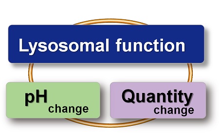
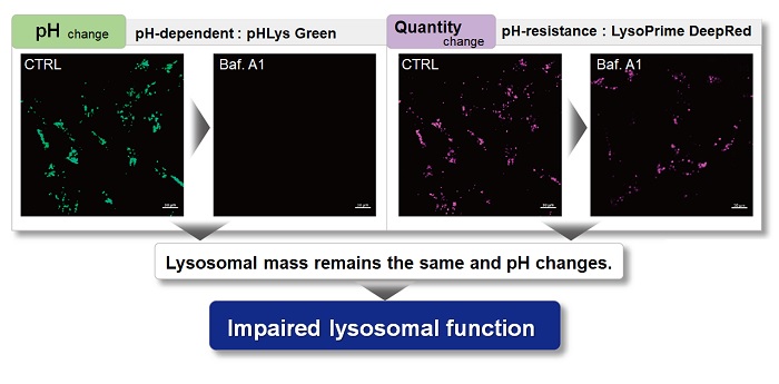
Lysosomal Function Analysis
With existing reagents, it was difficult to determine whether lysosomal mass or their function (pH) fluctuated because the discussion was based on changes in the fluorescence brightness of a single dye. This kit contains pHLys Green, which is highly specific to lysosomes and shows pH-dependent changes in fluorescence, and pH-resistant LysoPrime Deep Red. Using these two dyes, lysosomal pH and volume of the same sample can be measured for a detailed analysis of lysosomal function.
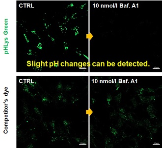
Accurate Measurement for Lysosomal pH changes
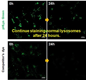
Existing lysosomal pH detection reagents have issues with dye localization, pH sensitivity, and retention. pHLys Green is a dye that solves these issues. The improved dye retention and localization enable detection of normal lysosomes, and the improved pH sensitivity enables detection of slight pH changes.
- 1. High sensitive pH detection
Comparison of pH response of cells treated with low concentrations of lysosomal acidification inhibitor Bafilomycin A1
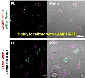
- 2. High specificity for lysosomes
Comparison of specificity for lysosomes using lysosomal marker protein LAMP1-GFP expressing cells
- 3. High retention in lysosomes
Comparison of intracellular retention
Product in Use:
- Lysosomal Acidic pH Detection Kit-Green/Deep Red
Related Product:
- pHLys Red- Lysosomal Acidic pH Detection
|


