Cellular Senescence: Review and Reagent Selection Guide

Science Note

Senescence at the Heart of Cancer and Neurodegeneration [Jun. 17, 2025] 

Cellular senescence is recognized as an important contributor to cancer development through its effects on the tumor microenvironment, and also to age-related dysfunction in both the peripheral and central nervous systems. This ScienceNote introduces recent findings that highlight the tumor-promoting role of senescent fibroblasts in breast cancer, the contribution of senescent neurons to age-associated pain, and the accumulation of senescent neurons through abnormal cell cycle activity in neurodegenerative conditions.

Senescent CAFs mediate immunosuppression and drive breast cancer progression (Cancer Discov., 2025)
Summary: This study identifies a subtype of senescent cancer-associated fibroblasts (senCAFs) in breast tumors that suppresses natural killer (NK) cell activity and promotes tumor growth. Eliminating these senCAFs enhances NK cell–mediated tumor killing and may offer a new therapeutic strategy to prevent breast cancer progression.

Highlighted technique: To examine how senescent CAF-derived ECM affects NK cell function, doxorubicin-treated mouse mammary fibroblasts were cultured for six days to deposit ECM in vitro. After cell removal, the ECM was used in NK cytotoxicity assays, with NK cells pre-incubated on the matrix before co-culture with labeled target cells.

 Related technique  SA-βGal Detection(Cell)

Aging and injury drive neuronal senescence in the dorsal root ganglia (Nature Neuroscience, 2025)
Summary: This study shows that peripheral sensory neurons (DRG neurons) undergo senescence with aging and nerve injury, leading to increased pain sensitivity via pro-inflammatory signaling and excitability. Eliminating these senescent neurons alleviated pain, suggesting a potential therapeutic strategy for age-related sensory dysfunction.

Highlighted technique: This study assessed DRG neuron senescence using multiple markers. Aging-related senescence was confirmed by increased SA-β-gal activity, and injury-induced senescence was evaluated by examining the upregulation of p16, p21, and SASP factors including IL-6.

 Related technique  SA-βGal Detection(Tissue)

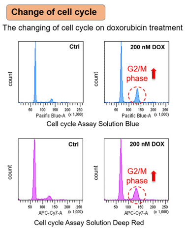
Neuronal cell cycle reentry events in the aging brain are more prevalent in neurodegeneration and lead to cellular senescence (PLoS Biol., 2024)
Summary: This study reveals that terminally differentiated neurons in the human brain, which are normally post-mitotic, can aberrantly re-enter the cell cycle under aging and neurodegenerative conditions. These neurons ultimately undergo senescence, with disease contexts like Alzheimer’s and Parkinson’s showing accumulation of proinflammatory and metabolically dysregulated senescent neurons.

Highlighted technique: To identify rare neurons that start dividing again, the researchers analyzed RNA data from individual cell nuclei collected from multiple human brain samples. They used a list of 350 genes related to the cell cycle and a scoring method to find excitatory neurons that are normally non-dividing but showed signs of abnormal cell cycle activity.

 Related technique  Cell Cycle Assay

Previous Science Note

Related Techniques (click to open/close)
Target Kit & Probes
Cellular senescence detection SPiDER-βGal for live-cell imaging or flow cytometry / microplate reader / tissue samples.
Blue cellular senescence detection dye for fixed cells,  SPiDER Blue
Cell Cycle Measurement Cell Cycle Assay Solution Blue / Deep Red
Mitochondrial membrane potential detection JC-1 MitoMP Detection Kit, MT-1 MitoMP Detection Kit
Total ROS detection Highly sensitive DCFH-DA or Photo-oxidation Resistant DCFH-DA
Oxygen Consumption Rate(OCR) Detection Extracellular OCR Plate Assay Kit
Apoptosis detection in multiple samples Annexin V Apoptosis Plate Assay Kit
Cell proliferation/ cytotoxicity assay Cell Counting Kit-8 and Cytotoxicity LDH Assay Kit-WST
 Application Note I  (click to open/close)
  > Senescent Cells Lose Mitochondrial Activity

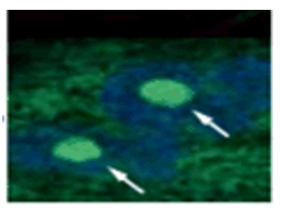
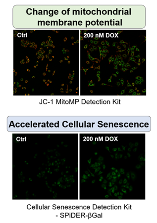
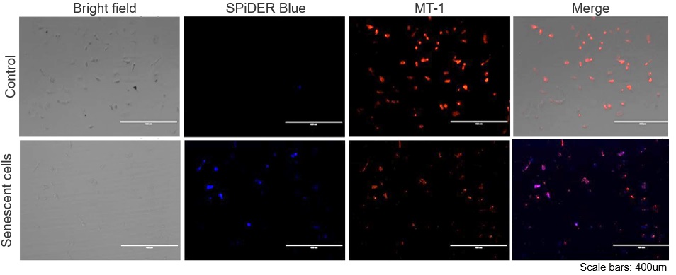
The senescent cell detection dye SPiDER Blue (SG07) and the mitochondrial membrane potential (MMP) dye MT-1 (MT13) were used to stain human microglial cells. Microscopy revealed that, compared to control cells, senescence-induced cells showed reduced MMP and increased SPiDER Blue fluorescence, reflecting elevated SA-β-Gal activity.

*This data was kindly provided by Dr. Supriya D. Mahajan, Department of Medicine, Jacobs School of Medicine & Biomedical Sciences.

1. Seed human microglia cells into a dish and incubate in an incubator set at 37 ℃ and equilibrated with 95% air and 5% CO2.
2. Dilute the MT-1 Dye (1:1000) in the cell culture medium.
3. Add MT-1 working solution to cells.
4. Incubate the cells for 30 minutes in an incubator set at 37 ℃ and equilibrated with 95% air and 5% CO2.
5. Discard the supernatant and wash the cells with HBSS twice.
6. Add 4% PFA/ PBS solution to the cells and incubate at room temperature for 30 minutes.
7. Discard the 4% PFA / PBS solution and wash the cells with PBS.
8. Add 15 µmol/l Spider Blue working solution and incubate at 37°C for 30 minutes.
9. Remove the working solution, and wash the cells with PBS.
10. Add Imaging Buffer solution and observe the cells under a fluorescence microscope.

 Application Note II (click to open/close)
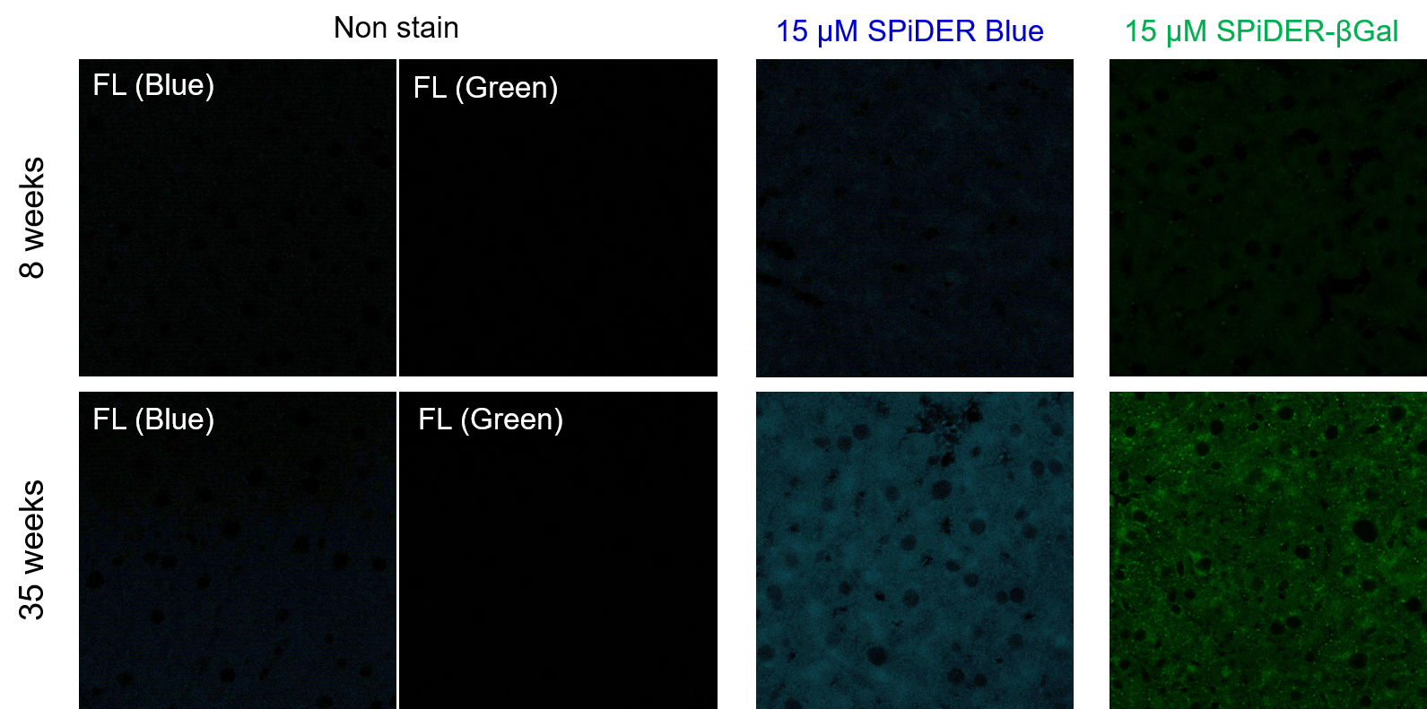
  > Increased Senescence in Aged Adipose Tissue

Frozen liver adipose tissue sections from 8-week-old and 35-week-old mice were stained with senescence detection probes SPiDER Blue (SG07) and SPiDER-βGal (SG02). Confocal microscopy revealed a marked increase in fluorescence intensity only in the 35-week-old samples, indicating an age-associated accumulation of senescent cells in older tissue.

1. 8-week-old and 35-week-old mouse liver adipose tissue (frozen sections) samples were prepared on glass slides. 
2. After washing once with PBS, 200 µl of 4% paraformaldehyde (PFA)/PBS solution was added and fixed at room temperature for 30 minutes. 
3. The supernatant was removed and washed once with PBS. 
4. Add 200 µl of 15 µM SPiDER Blue and 15 µM SPiDER-βGal prepared in Assay buffer and incubate at 37°C for 2 hours. 
5. The supernatant was removed and washed once with PBS. 
6. Add 1 drop of encapsulant (ProLong Glass Antifade Mountant, Thermo) and encapsulate with cover glass. 
7. Observed under a confocal laser microscope (40x magnification).

[Detection conditions]
SPiDER Blue: 405 nm (Ex), 400–500 nm (Em), 2.0%, 700V
SPiDER-βGal: 488 nm (Ex), 500–600 nm (Em), 1.0%, 600V


What is Senescence?

Lysosome
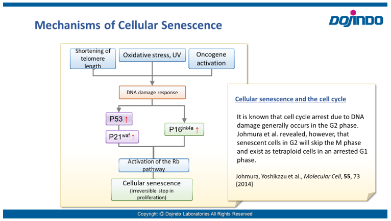
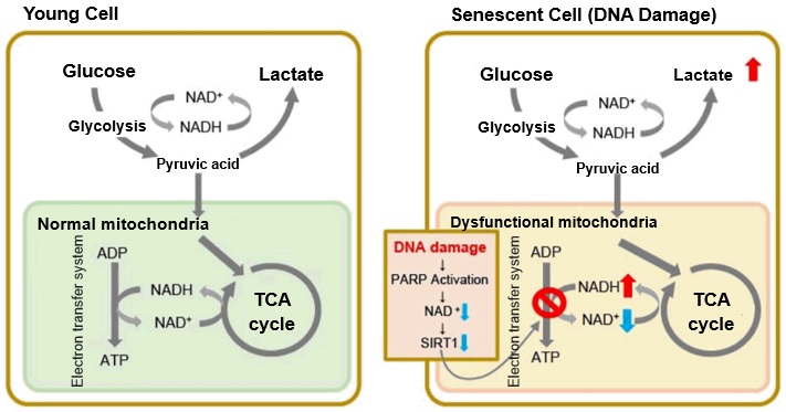
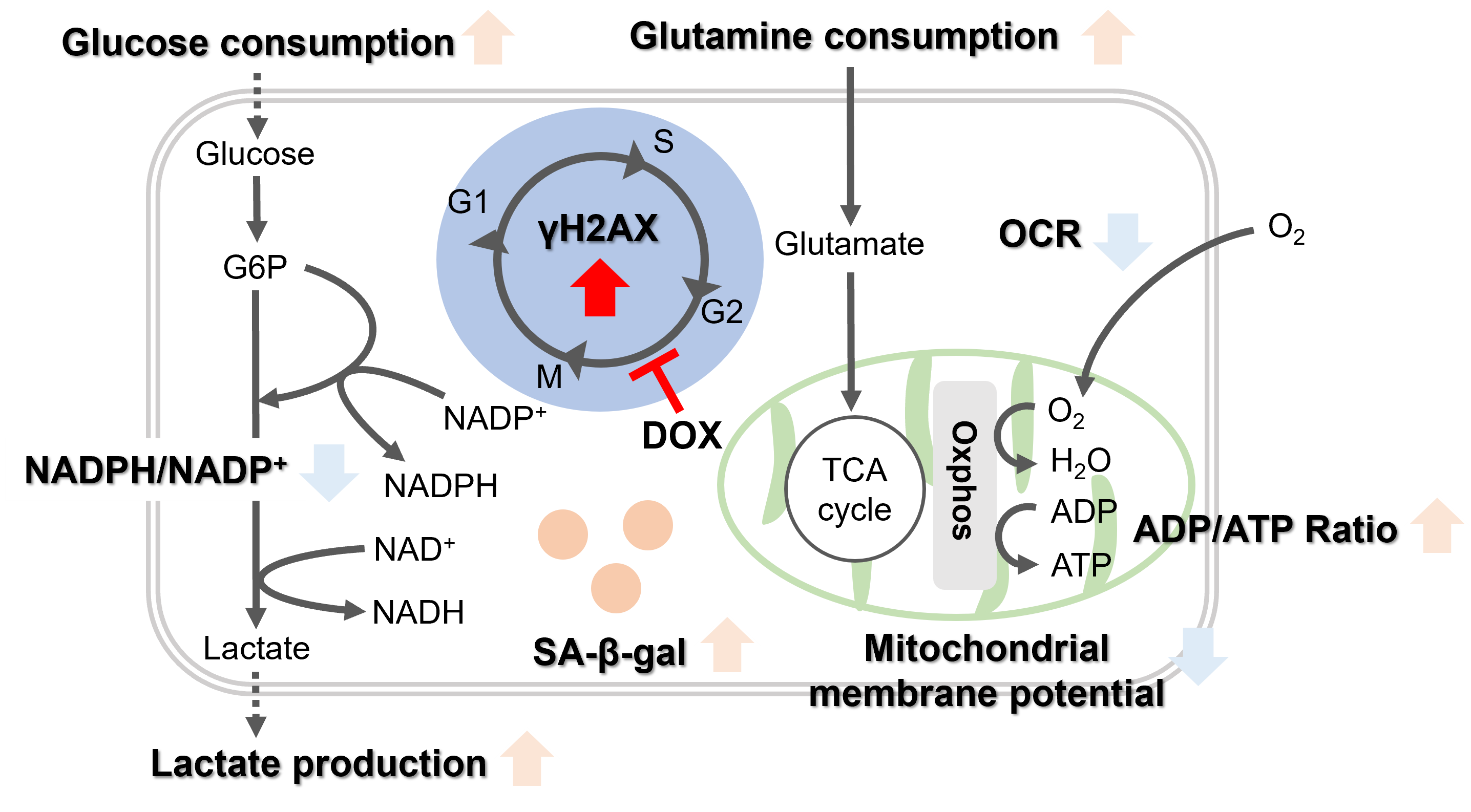
Senescence in cell biology refers to a state of permanent cell cycle arrest in response to stresses such as DNA damage or oncogene activation. Senescent cells can be identified by several molecular markers. Representative examples include p53, a transcription factor involved in the DNA damage response; p16, a cyclin-dependent kinase inhibitor that enforces cell cycle arrest; and senescence-associated β-galactosidase (SA-βgal). These cells resist apoptosis and ferroptosis and secrete inflammatory factors, collectively known as the senescence-associated secretory phenotype (SASP). Although senescence plays protective roles such as tumor suppression in early stages, the accumulation of these cells over time promotes chronic inflammation and contributes to several age-related diseases. As immune-mediated clearance of senescent cells declines with age, understanding their biology is crucial for the development of therapies targeting ageing and its associated diseases.

Assessing Cellular Senescence

Senescence
 Cellular senescence is controlled by various factors such as cell type and physiological conditions, such as oxidative stress. None of the individual biomarkers that have been identified so far have been deemed to be specific to senescent cells. Therefore, it is desirable to determine and confirm cellular senescence using multiple indicators.
 Common detection indicators for assessing cellular senescence include features related to cell cycle progression (DNA synthesis, p16/p21 expression, etc.), features related to morphology (of the cell, nucleus, nucleolus, etc.), SA-ß-Gal activity, DNA damage, oxidative stress (ROS), telomere length, inflammatory cytokines (senescence-associated secretory phenotype (SASP)), and more.


< Video Seminar >
“Recent Findings of Cellular Senescence Studies and Analysis Method”

Chapters:
0:00 What is Cellular Senescence ?
5:00 Senescence Studies and Drug discovery
12:30 Methods of Senescence Detection and Analysis

Reagent Selection Guide

Dojindo offers four types of kits and reagents that can be selected according to the evaluation method and purpose of cell senescence.

Product Cellular Senescence Detection Kit – SPiDER-ßGal, SPiDER Blue Cellular Senescence Plate Assay Kit – SPiDER-ßGal Cell Cycle Assay Solution Deep Red / Blue Nucleolus Bright Green / Red
Detection Fluorescence Fluorescence Fluorescence Fluorescence
Wavelength
(Ex/Em)
[SPiDER-ßGal]
Ex. 500–540 nm/Em. 530–570 nm
[SPiDER Blue]
Ex. 350-450 nm/Em. 400-500 nm
Ex. 535 nm / Em. 580 nm Deep Red: Ex. 633-647 nm /
Em. 780/60 nm
Blue: Ex. 405-407 nm /
Em. 450/50 nm
Green: Ex. 513 nm /
Em. 538 nm
Red: Ex. 537 nm /
Em. 605 nm
Target SA-ß-gal activity SA-ß-gal activity Nucleus Changes in the nucleolus
Detection
Method
Imaging, Flow cytometry
Substrate: SPiDER-ßGal, SPiDER Blue
Plate assay
Substrate: SPiDER-ßGal
Flow cytometry Imaging Detection of the nucleolus by RNA-staining reagent
Instrument Fluorescence microscope, FCM Fluorescence microplate reader FCM Fluorescence microscope
Sample SPiDER-ßGal: Live or fixed cells
SPiDER Blue: Fixed cells
(Tissue: some examples from published articles using SG02)
Live cells
(lysis of live cells)
Live cells, fixed cells Fixed cells
Best for Those who have difficulty quantifying data or performing multiple staining with X-gal Those who process multiple samples
Those who are evaluating senescent cells for the first time Small size package (20 tests) is available
Those who wish to evaluate using indicators other than SA-ß-Gal Those who wish to evaluate using indicators other than SA-ß-Gal
Examples of reports using nucleolus as an indicator are available on the product page
data
Item# SPiDER-ßGal: SG03(SG04)
SPiDER Blue: SG07
SG05 Deep Red: C548
Blue: C549
Green: N511
Red: N512


Indicators Related to Cellular Senescence

This correlation map shows the relationship between various intracellular indicators, resulting from cellular senescence. This information is based on currently available information. Please refer to the table with cited references below as reference for your experiments. The table lists the cell type, the method of senescence induction used, the senescence markers measured, and the variables affected by senescence in each reference for the map.

  Cell Senescence induction Senescence marker (s) Responding variable (s) Reference
IMR90
(Human pulmonary fibroblasts)
Several passages in culture SA-ß-Gal, p16, p21, Nucleosome hypertrophy Expression of SETD8↓,  H4K20me1↓, oxidative phosphorylation↑, ribosome synthesis↑ H. Tanaka, S. Takebayashi, A. Sakamoto, N. Saitoh, S. Hino and M. Nakao, “The SETD8/PR-Set7 Methyltransferase Functions as a Barrier to Prevent Senescence-Associated Metabolic Remodeling.”Cell Reports2017, 18(9), 2148.
Inhibition of SETD8
(Methyltransferase)
Oxidative phosphorylation↑,  ribosome synthesis↑
Senescent mouse satellite cell
eletal muscle progenitor cells)
SA-ß-Gal, p16 Autophagy activity↓, ROS↑, mitochondrial membrane potential L. Garcia-Prat, M. Martinez-Vicente and P. Munoz-Canoves, “Autophagy: a decisive process for stemness”Oncotarget2016, 7(11), 12286.
Atg7 knockout mouse
(Satellite cells)
Autophagy inhibition SA-ß-Gal, P15, p16, p21, γ-H2AX ROS↑, mitochondrial membrane potential
Rat fibroblast model of type 2 diabetes SA-ß-Gal, p21, p53, γ-H2AX NADP+/ NADPH↓(resistance to oxidative stress↓), NADPH oxidase↑(ROS↑) M. Bitar, S. Abdel-Halim and F. Al-Mulla, “Caveolin-1/PTRF upregulation constitutes a mechanism for mediating p53-induced cellular senescence: implications for evidence-based therapy of delayed wound healing in diabetes”Am J Physiol Endocrinol Metab.2013, 305(8), E951.
IMR90
(Human pulmonary fibroblasts)
Ethidium bromide (inhibition of mtDNA) + pyruvate deficiency SA-ß-Gal NAD+/NADH C. Wiley, M. Velarde, P. Lecot, A. Gerencser, E. Verdin, J. Campisi, et. al., “Mitochondrial Dysfunction Induces Senescence with a Distinct Secretory Phenotype”Cell Metab., 2016, 23(2), 303.
MDA-MB-231
(Human breast cancer cells)
X-ray irradiation + inhibition of cell cycle-related factor (securin) expression SA-ß-Gal Lactate↑, LDH activity↑, (glycolysis↑) E. Liao, Y. Hsu, Q. Chuah, Y. Lee, J. Hu, T. Huang, P-M Yang & S-J Chiu, “Radiation induces senescence and a bystander effect through metabolic alterations.”Cell Death Dis., 2014, 5, e1255.
MEF
(Mouse Embryonic Fibroblast)
Overexpression of oncogenes,several passages in culture, transcription factor overexpression(E2F1) SA-ß-Gal, p16, p21, Nucleosome hypertrophy Ribosome RNA↑, p53↑ K. Nishimura, T. Kumazawa, T. Kuroda, A. Murayama, J. Yanagisawa and K. Kimura, “Perturbation of Ribosome Biogenesis Drives Cells into Senescence through 5S RNP-Mediated p53 Activation”Cell Rep2015, 10(8), 1310.
Mouse tail fibroblast 2 months old, 22 months old, p16 knockout (22 months old) SA-ß-Gal, p14, p16 NAD+↓, SIRT3↓ M. J. Son, Y. Kwon, T. Son and Y. S. Cho, “Restoration of Mitochondrial NAD+ Levels Delays Stem Cell Senescence and Facilitates Reprogramming of Aged Somatic Cells”Stem Cells2016, 34(12), 2840.


Accumulation of Lipid Peroxides and Their Connection to Cellular Senescence and Mitochondria

Lipotoxicity is caused by intracellular lipid accumulation and is indicative of mitochondrial disfunction. Lipotoxicity accelerates the degenerative process of cellular senescence, influencing cancer development.

References

1. Clara, C. al., “Mitochondria: Are they causal players in cellular senescence?”, Biochimica et Biophysica Acta – Bioenergetics20151847(11), 1373-1379.

2. Huizhen, Z. et al., “Lipidomics reveals carnitine palmitoyltransferase 1C protects cancer cells from lipotoxicity and senescence”, Journal of Pharmaceutical Analysis2020.

3. Xiaojuan, H. et al., “Astrocyte Senescence and Alzheimer’s Disease: A Review”, Front. Aging Neurosci.2020.

4. Borén, J. et al., “Apoptosis-induced mitochondrial dysfunction causes cytoplasmic lipid droplet formation”, Cell Death Differ201219(9), 1561-1570.

5. Na, L. et al., “Aging and stress induced β cell senescence and its implication in diabetes development”, Aging (Albany NY)201911(21), 9947–9959.

Cell Cycle Arrest

 Irreversible cell cycle arrest is one of the phenomena that characterize cellular senescence. p16, p21, p53, and pRB (phosphorylated retinoblastoma protein) are known as representative protein markers. The activation/upregulation of these proteins are used as indicators of cellular senescence. These marker proteins are known to be tumor suppressors and regulate the cell cycle mainly through two pathways (p16Ink4a-RB and p53-p21CIP1).

Doxorubicin (DOX) is known as an anticancer drug that acts in the G2/M phase of the cell cycle to arrest cell proliferation and induce cellular senescence (see the figure below in center). Below are the results of an experiment in which DOX was added to A549 cells. As a result, changes in SA-ß-Gal expression, cell cycle progression, and mitochondrial membrane potential were observed.

Changes in Intracellular Metabolism

In aged cells, due to mitochondrial dysfunction, ATP is primarily generated through the anaerobic glycolysis pathway, leading to an increase in lactate production2). DNA damage is one of the causes of mitochondrial dysfunction in cellular aging. The accumulation of DNA damage activates DNA repair mechanisms and increases NAD+ consumption. The decrease in NAD+ levels reduces SIRT1 activity, an important factor in maintaining mitochondrial function, leading to impaired mitochondrial function (inhibition of electron transfer → ATP production / reduction of NAD+ levels)1),3).

Reference:

1. J. Wu, Z. Jin, H. Zheng and L. Yan, “Sources and implications of NADH/NAD+redox imbalance in diabetes and its complications”, Diabetes Metab. Syndr. Obes., 2016, 9, 145

2. Z. Feng, R. W. Hanson, N. A. Berger and A. Trubitsyn, “Reprogramming of energy metabolism as a driver of aging”, Oncotarget., 2016, 7(13), 15410.

3. S. Imai and L. Guarente, “NAD+ and sirtuins in aging and disease”, Trends in Cell Biology, 2014, 24(8), 464.


Oxidative stress & accelerated aging:
SA-β-gal
Impairment of mitochondrial function:
③ Mitochondrial membrane potential
④ Oxygen consumption rate (OCR)

⑤ ADP/ATP ratio

Upregulation of glycolysis pathway and glutamine metabolism
⑥ Glucose consumption
⑦ Lactate production
⑧ Glutamine consumption
Reduction in antioxidant capacity:
⑨ NADPH/NADP+ ratio
DNA repair mechanisms:

⑩ NAD+/NADH Ratio


Product Classification

Product Classification