Si-DMA for Mitochondrial Singlet Oxygen Imaging

Mitochondrial Singlet Oxygen Imaging
-
Product codeMT05 Si-DMA for Mitochondrial Singlet Oxygen Imaging
-
CAS No.1621598-01-3
-
Chemical nameN-[10-(9,10-DimethylanthraceN-2-yl)-7-(dimethylamino)-5,5-dimethyldibenzo[b,e]siliN-3(5H)-ylidene]-N-methylmethanaminium, chloride
-
MWC35H37ClN2Si=549.22
| Unit size | Price | Item Code |
|---|---|---|
| 2 μg | $315.00 | MT05-10 |
Description
Singlet oxygen (1O2) is one of the Reactive Oxygen Species (ROS). 1O2 is known to be a cause of spots and wrinkles of the skin due to its very strong oxidizing potential. In the field of cancer research, 1O2 is of a particular importance because of its key role in photodynamic therapy (PDT), an emerging anticancer treatment using photoirradiation and photosensitizers. For the detection of 1O2, the existing singlet oxygen fluorescence detection reagent cannot be used in living cells because of its cell membrane impermeability.
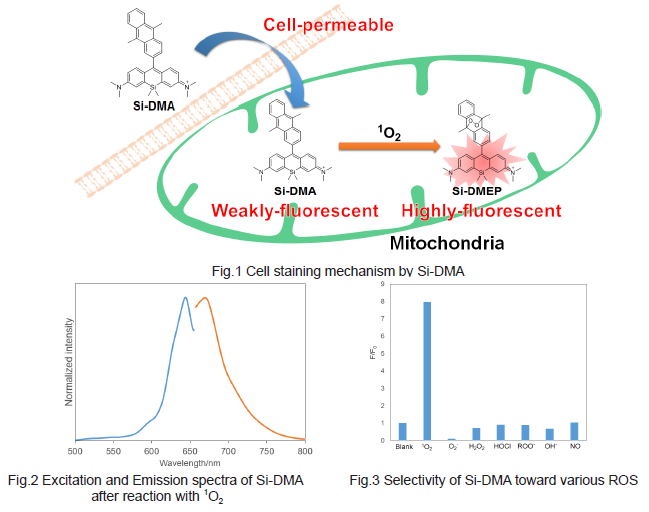
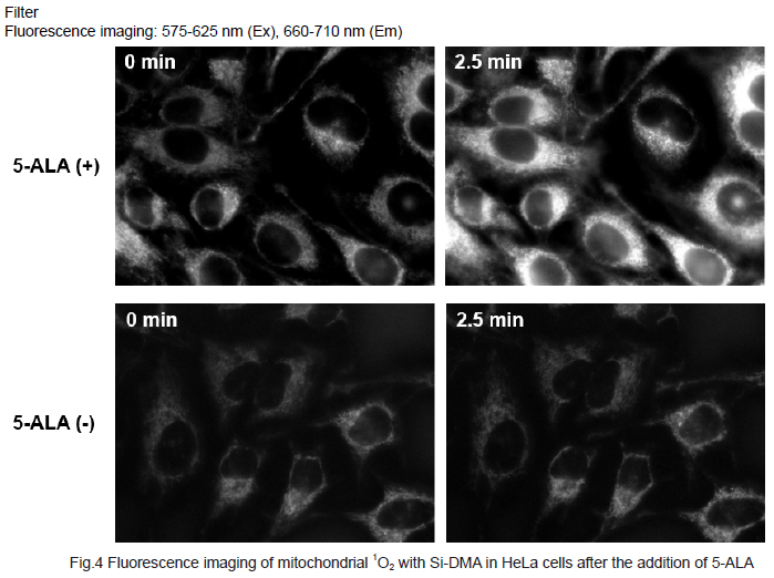
Majima et al. synthesized a new far-red fluorescence probe composed of silicon-containing rhodamine and anthracene moieties, namely Si-DMA, as a chromophore and a 1O2 reactive site, respectively. In the presence of 1O2, fluorescence of Si-DMA increases 17 times due to endoperoxide formation at the anthracene moiety(1),(2). Among seven different ROS, Si-DMA is able to selectively detect the 1O2 (Fig. 3). In addition, Si-DMA is able to visualize the real-time generation of 1O2 from protoporphyrin IX in mitochondria with 5-aminolevulinic acid (5-ALA), a precursor of heme(Fig. 4).

| Developer | Dojindo Molecular Technologies, Inc. |
|---|
Manual
Technical info
Fluorescence microscopic detection of 1O2 in HeLa cells after added 5-aminolevulinic acid (5-ALA)
1. HeLa cells (2.4×10⁵ cells/ml, Hanks’ HEPES buffer 200 μl) were seeded on a μ-slide 8 well (Ibidi) and cultured at 37oC in a 5%CO2 incubator overnight.
2. Cells were washed with 200 μl Hanks’ HEPES buffer twice.
3. 150 μg/ml of 5-ALA in Hanks’ HEPES buffer was added to the μ-slide and cultured at 37oC in a 5%CO2 incubator for 4 hours.
4. Cells were washed with 200 μl Hanks’ HEPES buffer twice.
5. 200 μl of Si-DMA working solution (40 nmol/L) was added and cultured at 37oC in a 5%CO2 incubator for 45 minutes.
6. Cells were washed with 200 μl Hanks’ HEPES buffer twice.
7. 200 μl Hanks’ HEPES buffer was added and observe the cells under a fluorescence microscope.

References
1) S. Kim, T. Tachikawa, M. Fujitsuka, T. Majima, “Far-Red Fluorescence Probe for Monitoring Singlet Oxygen during Photodyanamic Therapy”, J. Am. Chem. Soc., 2014, 136 (33), 11707.
2) S. Bekeschus, A. Mueller, U. Gaipl, KD. Weltmann, "Physical plasma elicits immunogenic cancer cell death and mitochondrial singlet oxygen", TRPMS., 2017, 99, DOI:10.1109/TRPMS.2017.2766027.
Handling and storage condition
| Appearance: | Blue to blueish green solid |
|---|---|
| Purity (HPLC): | ≧ 90.0 % |
| NMR spectrum: | Authentic |
| -20°C, Protect from light |


